
Introduction to Course
Austin Lim
Additional Videos and Readings
This video first offers a historical overview of some of the major milestones that lead to our current understanding of the brain’s control over behavior. Then, some of the fundamental questions asked by behavioral neuroscientists are presented, which all involve the study of neural correlates, or specific brain regions whose activation is responsible for a given function. Next, prominent methods used to answer those questions are reviewed for both human and animal subjects, such as operant conditioning and functional neuroimaging. Finally, experimental applications of these techniques are presented, including animal training using a Skinner box, and the use of electroencephalography to investigate human neurological disease.
Video– Ted Talks; What is so special about the human brain? by Suzana Herculano-Houzel
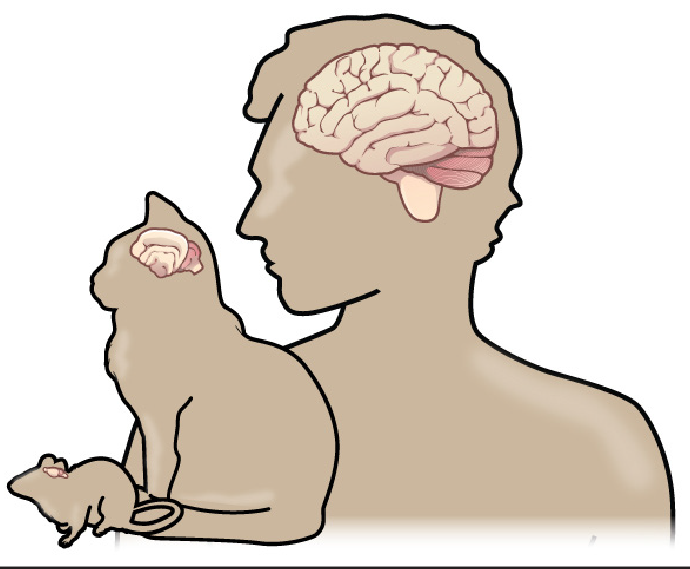
Suzana Herculano-Houzel discusses what distinguishes human brains from other animals, giving an overview of her research to count the number of neurons in different types of brains. Her research shows that humans have a remarkable number of neurons in the cerebral cortex, made possibly by the invention of cooking, which allowed us to consume and metabolize enough energy to fuel our brains in a way that other primates could not.
What is Neuroscience?
In short, neuroscience is the study of the nervous system, the collection of nerve cells that interpret all sorts of information, which allows the body to coordinate activity in response to the environment.
The study of neuroscience has taught us that the brain is a complicated organ with several connection routes, both between different bodily organs and within itself. Some of those connections communicate information down towards the body, such as signals that allow us to control the movements of our muscles, or to change the activity of our internal organs. Other connections ascend into the brain, conveying all sorts of information from the world around us into a representation of our surroundings. Still, other routes communicate between brain areas, such as when the sudden detection of a threat passes through our visual system and turns into a “get ready” signal that then prepares the rest of our body for conflict. Because of this complex system of communication, the nervous system can be thought of as a series of highways and roads that connect different cities (organs.)
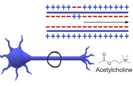
The nervous system conveys all of these different types of information using a combination of electrical and chemical signals. The main active cellular units of the nervous system, the , are highly sensitive to changes in their environment. Similar in the way that computers do all their work using a highly coordinated binary signal of 0s and 1s, the electrical output of many neurons is an all-or-nothing response called an . A wide variety of chemicals called is responsible for passing information between neurons.
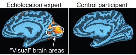
The brain is the main computational powerhouse of the body, much more complex and intricate than any artificially created system thus far. Estimations of the computational power of the brain suggest that we can handle somewhere around 10^28 operations per second, processing power that is many orders of magnitude faster than any supercomputer to date. While it’s true that computers can do large, mathematical calculations that (most) humans can’t, the real strength of our brain is its flexibility: brains are capable of changing and adapting to a wide variety of circumstances. Blind people use their visual areas of the brain while echolocating , stroke survivors can regain lost motor functions using the unaffected brain circuits, and babies can effortlessly learn two languages simultaneously in a bilingual household.
The brain is also responsible for the most abstract and unique human functions, such as the origin of consciousness, the place where our thoughts, fears, and desires are born, and the endless creativity of our species. All the musical works of Mozart, the literary genius of Shakespeare, and the philosophical theories of Aristotle were produced through some complex interaction of neurons that we may never understand.
How do we learn about Neuroscience?
Generally speaking, there are three main research designs that have been used- and will likely continue to be used – to learn about the brain.
Experimental design
The gold standard in science is the use of . In an experiment, the scientist uses a stepwise process of developing a research question and hypothesis, then answering that question by performing tests. The main goal of an experiment is to establish a causal relationship between one factor that is being changed, the , and the factor that is influenced, the . A well-designed experiment has variables that are carefully controlled, which minimizes the influence of extraneous variables, often called . The influence of confounding variables can be eliminated by comparing the experimental group with a , a group that is as similar as possible in every way except for the manipulation of the independent variable. Importantly, subjects or patients are generally assigned to the experimental or control group at random.
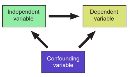
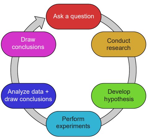
For example, consider the research question: “Does studying more increase performance on exams?” Here, the independent variable is the number of hours studied, while the dependent variable is the grade on the exam. A potential confounding variable could be the number of hours slept before the exam, since poor sleep causes poor memory recall performance, and students may choose to study instead of sleep. To eliminate this confounding variable, it would be good to only compare grades for the students who slept for roughly the same amount of time. A control group in this experiment would be a group of students who are given the test without the opportunity to study. Ideally, these students will be as similar to the experimental group as possible: roughly the same age, gender distribution, educational history, and so on.
The strength of a well-designed experiment is that it establishes : a change in the independent variable causes a change in the dependent variable. Because of this, assuming that the sample population is (the distribution of the characteristics in the sample is proportionally similar to the distribution in the total population,) experiments allow us to extrapolate findings to a larger population.
Patients who participate in an experiment are often placed into an artificial environment or unnatural circumstances, which can affect their performance. For example, imagine you were participating in the “studying / test score experiment.” Having the added pressure of knowing you were in a study could cause you to perform worse. Alternatively, knowing that you are in the experimental group might cause you to focus more intently during your study time, which could increase the group’s average. To ameliorate this unintended effect, it’s important for participants in the experimental group and the control group to be subjected to the exact same circumstances.
Observational study
A second way to gain scientific information is through an observational study, one type of which is a . These studies do not benefit from separation of patients into groups randomly, and therefore may have several uncontrolled variables. Quasiexperimental studies are usually done when conducting an experimental study may be too impractical or otherwise unethical.
An example of this type of study might aim to answer the question “Do people with traumatic brain injury have worse hand-eye coordination?” Performing an experiment with this question would be wildly unethical, since you cannot intentionally give your patients head injuries (It would also be challenging to find people willing to get hit on the head for science!). Instead of conducting this unethical experiment, you could answer this question with a quasiexperimental study. In the study, there would be two different populations of patients, an experimental group containing people who have already been diagnosed with head injury, and a control group that is demographically similar, but without a diagnosis of head injury. You could then ask both groups to perform a hand-eye coordination task and see ifthere are differences in their performance.
The weakness here is that there is no true randomness in the groups, thus the influence of confounding variables may affect our interpretation of the data. If the head injury group performed worse on the hand-eye coordination task, you would be able to demonstrate correlation, but not causation, which may present as a “chicken or the egg” problem. Perhaps the head injury group already had poor hand-eye coordination, which led to a car / workplace / sports accident, resulting in their head injury. In this example, we cannot conclude whether the brain injury directly causes a worsening of hand-eye coordination.
Case Study

 A third strategy is the , a highly detailed description of a single patient and their condition. A case study documents the details regarding a specific deficit or enhancement, and is an opportunity to examine individuals with very rare conditions, which are useful for informing about the functions of different brain structures. Examining millions of healthy people may not give us the same insight as studying just one person with a specific injury or disorder. For example , case studies have been instrumental in teaching us about the brain structures involved with memory (Patient HM; chapter 13), language (Patient Tan; chapter 14), and fear processing (Patient SM; chapter 15).
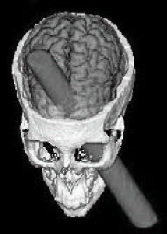
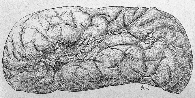
Perhaps the most famous case study in all of neuroscience is the 1848 story of the railroad worker Phineas Gage. An unfortunate workplace accident left him with significant brain damage, largely to his frontal lobe. The subsequent changes in his personality taught us that one of the functions of this area of the brain is regulating our inhibitions (chapter 2).
Like a quasi-experimental study, case studies only show correlation, not causation. It is difficult to generalize the findings from a case study to the population at large. Usually, case studies are descriptions of nearly-one-of-a-kind individuals: A man with memory deficits in response to his brain being punctured by a toy fencing sword, a woman who had never experienced fear in her life, or a man who became overwhelmingly light-hearted and joyful after recovering from being shot in the head.
Case studies can be helpful for the development of hypotheses that can later be tested experimentally. For example, consider Patient HM, the man who had his left and right hippocampus surgically removed and couldn’t create certain types of memory. A research question based on this case study might be “Is the hippocampus needed for the creation of navigational memory?” Then, an experimental study could be performed in rodents, where we surgically remove the hippocampus (experimental group) or a different part of the brain (control group) and see how well the rodents perform on a memory task.
What Neuroscience is NOT
As complex as the brain is, naturally misconceptions make their way into popular culture. It’s valuable to address these myths about neuroscience and explain the evidence that refutes these statements.
 “We only use 10% of our brain.”

This wildly-inaccurate statistic has been the foundation for several fictional movies, TV shows, and books. The truth is that we use every part of the brain, and most of our brain is active most of the time – just not at the same time. Neurologist V.S. Ramachandran uses a great analogy to describe the fallacy of this myth: does a traffic light only use 33% of its lights? A properly functioning traffic light will use all three lights at very precise times. The activity of the brain is closely regulated by multiple mechanisms which prevent unusual electrical activity. In fact, if too many cells were active at the wrong times, just like a traffic light showing both green and red, chaos ensues – one cause of seizures is excessive neural activity.
 “Forming memories causes new neurons to be born.”
Another misconception is the idea that each new cell in our brain represents a new memory. While we are far from understanding the process of exactly how memories are formed in the brain, we do have a few clues. Most likely, memories are stored at the sites of close contact between neurons, called . Changes in in ways neurons connect and communicate with one another is likely the mechanism behind how memories are formed and stored, rather than the creation of new neurons.
Even though the process of cell reproduction is halted in the majority of adult neurons, we are still capable of new neuronal growth, a process called . A few brain areas in particular, like the hippocampus (used in learning and memory functions; chapter 13) and the olfactory epithelium (used for smelling; chapter 9), do exhibit frequent birth and death of new neurons.
 “The brain cannot repair itself.”
If neurons aren’t being replaced in adulthood, then how do people spontaneously recover from neurological injuries like a stroke?
One of the most amazing features of the brain is the phenomenon of , the ability to change over time. Even if critical brain areas are damaged, it is theorized that the brain learns how to “rewire itself,” essentially figuring out how to carry out these functions without using the damaged connections.
Unfortunately, there are some conditions that are , meaning that their symptoms get progressively worse over time. Many of these disorders, like Parkinson’s disease (chapter 10) and Alzheimer’s disease (chapter 13), currently don’t have any simple cures or treatments that don’t carry risks and side effects. For people with these conditions, there isn’t strong evidence that the brain can recover from the destruction caused by these diseases.
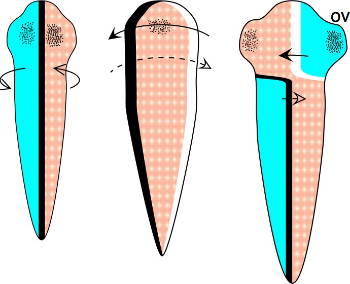
Fascinatingly, we do have one strange quirk about signaling between the brain and the rest of the body: signaling pathways from the left brain crosses over to communicate with the right half of the body, and vice versa. This organization is an unintended consequence o f evolution, and is one of the major distinguishing features of the vertebrate brain.
 “If you are analytical, you are left brain dominant, but if you are creative, you are right brain dominant.”
A common misconception is that the two hemispheres of the brain are responsible for wildly different functions. The truth is that nearly every function that the left half of the brain can do, the right half can do just as well, and vice versa. Sensory information, voluntary control of the muscles, memories, and many other behaviors can be performed equally well by both the left and right halves of the brain.
A major exception to the “left vs. right” component is the processing and production of language. For some reason unknown to scientists, these functions are heavily lateralized in the left hemisphere for most people (chapter 14).
Neuroscience is ever changing

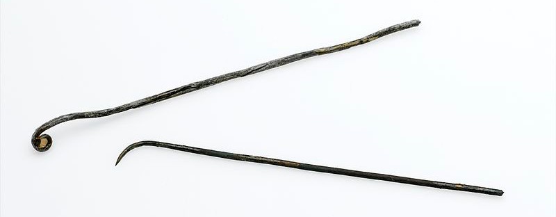
One of the most exciting and satisfying aspects of modern science is the rapidity of new discoveries the field. New findings are often communicated by publishing academic studies in scientific journals. More neuroscience studies were published between 2015 and 2020 than in the previous seventy years! But, advancements in neuroscience haven’t always moved so quickly. The ritualistic funerary rites of the ancient Egyptians around 2500 BCE provide a glimpse into how humankind’s understanding of the brain has changed over time. When important Egyptians died, major organs including their stomach, lungs, and liver, were removed and stored in canopic jars in preparation for immortality in the afterlife. The fate of the brain, however, was much messier: Using a pair of sticks up the nose, the brain was blended up into a mush and flushed out of the skull using palm wine. The brain, apparently, wasn’t needed for the afterlife.
Around 2,000 years later, ancient Greek physicians had a different understanding of the function of the brain. Aristotle developed a theory that the heart was the seat of the soul, and that blood was the life force that dictated a person’s behavior. When a person was “hot blooded,” they acted impulsively with no regard for consequences. In his view, the function of the brain was to cool the blood as the blood passed through it, which calmed the temper.
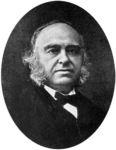
For the hundreds of years that followed, physicians attempted to correlate behaviors with changes in the brain. In the mid 1800s, Paul Broca was one of the first to suggest that specific areas of the brain were responsible for carrying out specific functions, which came to be called . Much evidence favors this line of thinking, such as the idea that language comprehension starts in a small patch of cells in the left hemisphere (Chapter 14), perception of faces relies on a set of cells at the base of the brain (Chapter 7), and balance and motor coordination depends on the cerebellum (Chapter 10). On the other hand, the opposing view, called the , suggests that behavioral functions require activation of cells across several different areas of the brain. Complex behaviors such as emotion, consciousness, or (the act of generating knowledge through a combination of senses, memories, and thoughts) require coordinated action across distinct brain areas. Most likely, some behaviors are more localized than others, but still rely on signals from across many other brain areas. As with most fields of biology, absolutes are rare in neuroscience.
While anatomists and physicians tried to define the gross anatomical workings of the brain, they missed out on a layer of understanding at the level of cells until microscopy was widely adopted by the scientific community. In the early 1900s, a heated debate between two anatomists, Camillo Golgi and Santiago Ramon y Cajal, prompted researchers to look more closely at the neurons. Through careful drawings of their observations, they concluded that neurons had different shapes and therefore carried out different functions. This microscopic level analysis laid the foundation for understanding the cells that make up the nervous system and the way they communicate with one another (Chapter 2).
Today, we have a clearer understanding of the function of the brain, largely due to the advancements brought to us by a better understanding of animal biology and new technology. In 1954, the was aimed at the space between neurons for the first time, allowing us to see a tiny anatomical component about 20 nanometers across – a thousand times smaller than the width of a human hair (Chapter 5). A medical diagnostic tool, the functional magnetic resonance imaging device (fMRI), made its debut to the neuroscience world in 1991, which allowed us to visualize brain activity while a person is actively engaged in behaviors, such as a decision making task, or while observing visual stimuli (Chapter 6). Today, much excitement revolves around visualization strategies like , a method to render an entire brain transparent, which helps us to map out the nature of the connections that span the nervous system.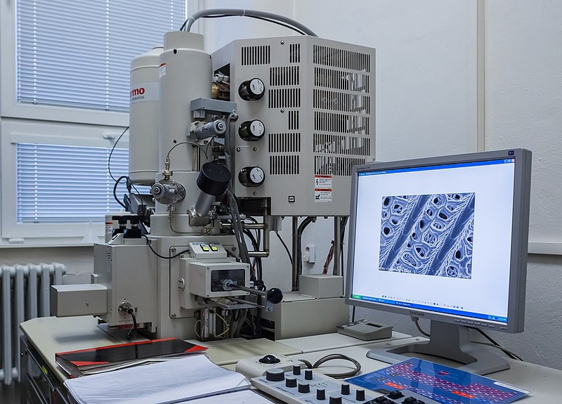
The ever-changing landscape of scientific inquiry presents a challenge. Our current understanding of the brain, as described here, is only a snapshot along the timeline of scientific discoveries. As we look to the future, many new discoveries will continue to reinforce what knowledge we have already amassed. But some discoveries, with help from not-yet-invented technology, will push the frontiers of knowledge and find compelling evidence against long- standing accepted theories in the field , prompting a shift in the paradigm.
Neuroscience is an integrative field of study
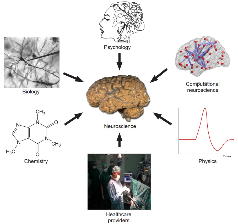
Realistically, our modern understanding of “neuroscience” is a combination of several academic disciplines, all using their strengths to understand some aspect of the nervous system. Because of this integrative nature, it is possible to study neuroscience from many different perspectives, each of them more fitting for answering different types of questions . These “angles” of analysis are described below.
At the root of the study is biology. Whenever you are studying living processes, such as learning, visual perception, or consciousness, you dip into the realm of biology. The broad field of biology can be subdivided into smaller, more precise categories. Molecular neurobiologists study proteins and gene regulation, cellular neurobiologists examine how networks of neurons communicate with one another, and cognitive neuroscientists study the underlying causes of behaviors. Understanding neuroscience involves genetics, such as the autosomal dominant neurodegenerative condition, Huntington’s disease (Chapter 10.) Other biological subdisciplines such as ecology and evolution are also considered in neuroscience as well, such as the parasite Toxoplasma, which changes an animal’s response to fearful stimuli, allowing the organism to reproduce as it moves through different species in the food web (Chapter 15.)
Psychology provided the earliest explanations about the brain and ideas about the origin of the mind. Some questions in this field branched off from philosophy, as people began thinking about the “,” the discussion that centered around the question if a function as complex as consciousness could result from activity of a clump of cells.
Psychologists also wondered whether parts of the brain in isolation have different properties than when those parts are working together. This property, called , is the idea that the whole is greater than the sum of its parts. Psychologists examine neuroscience from a top- down view, aiming questions at understanding the whole organism before looking at smaller components of the organism (compare this with biological approaches, often a bottom-up view that starts at the level of cells or molecules.)
Chemistry is a strong influencer of nervous system function – just ask anyone who forgot their morning cup of coffee! We use a variety of (originating from within the body) chemicals that act as signaling molecules, allowing communication between cells. These chemicals exist in many different structures, which determine their function. Some are acidic while others are basic; some are polar, others are fat soluble, and some are even gases (chapter 5). The nervous system is also highly sensitive to influence by chemicals (meaning they originate from outside the body), such as caffeine and cocaine (Chapter 11).
Many principles of physics can be observed through the functioning of neurons. For example, neurons maintain a negative electrical charge, usually measured on the scale of tens of millivolts (a millivolt is a thousandth of a volt.) The main way for neurons to send signals depends on a temporary change in this voltage; this signal is called an . This change in voltage is brought on by the movement of charged ions across the cell membrane, and they closely follow the rules of magnetism: opposite charges attract while like charges repel (Chapter 4).
The field of has grown from the use of mathematical modeling to describe or predict some aspect of the nervous system. If our current estimates are correct, we have around 86 billion neurons in the brain, a number so large that it is difficult to conceptualize. It would be nearly impossible to understand that many components of a system without taking advantage of the sheer mathematical strength of a computer.
Healthcare providers, like neurologists and psychiatrists, work from a different angle. They coordinate closely with researchers to apply scientific knowledge from the field or laboratory to treat patients, thus using biological principles as therapies. For example, neurologist Dr . Oliver
Sacks used his knowledge of the dopamine neurotransmitter system to treat patients with a paralysis-like condition in the 1960s, leading to the development of levodopa treatment for Parkinson’s disease (chapter 5). Other healthcare providers use imaging strategies like a CT scan to assess the extent of a head injury or the location of a brain tumor, while an EEG can be helpful for the diagnosis of epilepsy (Chapter 6).
Engineers help develop the tools needed to understand questions in neuroscience, such as the patch clamp rig or electron microscope, highly specialized pieces of lab equipment. They also work closely with healthcare providers to translate science into therapy, such as the deep brain stimulator devices for the treatment of conditions such as Parkinson’s disease.
Collectively, all the people who participate in neuroscience in some way are united by their interest in the workings of the body. Because of the overwhelming complexity of the nervous system, there are many questions still unanswered. The continual appearance of new questions in neuroscience keeps us wondering, inspires curiosity, and promises a multitude of fascinating career paths for centuries to come.
Chapter 3: Cellular Anatomy of the Nervous System
For the majority of human history, the only way we were able to study the structure of the brain was with crude, butcher-like methods. Wait for a person to die, saw a giant hole in the top of the skull, take the brain out, and slice it into pieces to see if there was some correlation between the way the brain looks and the way they died.
With these methods, only major changes in gross anatomy could be observed, such as that resulting from severe birth defects or trauma. Brain analysis methods were enhanced by the scientific adoption of light microscopy. Naturalists in the mid 1600s such as Antonie van Leeuwenhoek, Jan Swammerdam, and Robert Hooke began looking at biological substances up close, and the brain proved to be a complex and interesting sample of tissue.
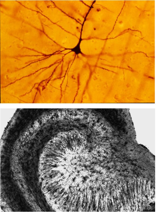
A major advancement in the study of neuronal morphology came about in the late 1800s. The Italian anatomist and biologist Camillo Golgi identified a shortcoming with the cellular analysis techniques of the time: structures in the CNS were impossible to distinguish from one another. The cells in the brain were so densely packed together, that it became difficult to identify which cellular material belonged to which cell. Golgi came up with a new technique using a silver compound that caused the silver to precipitate inside the cell membranes. However, not every cell took up the silver. Instead, only a small fraction of neurons, maybe 1% or even less, were completely stained in black, which stood out remarkably well against the light yellow background of the surrounding tissue. This reaction, initially called the “black reaction,” is now known as a “” (Despite being more than a hundred years old, we currently don’t know the mechanism by which the silver stain is taken up into the neurons, or what determines why certain cells take the stain and others don’t.) Because of the great contrast between cell and background, every single part of the neuron was completely filled, allowing Golgi to do drawings of the morphology of this nervous tissue. Based on his staining results, Golgi supported the idea that the parts of the nervous system are all one very large, physically connected network. This idea was known as the .
About 10 years later, the Spanish neuroanatomist Santiago Ramon y Cajal repeated some of Golgi’s staining experiments with other sections of nervous tissue. Looking at similar darkly-filled neurons, Cajal arrived at a different conclusion: the nervous system is not a giant net, but rather a series of individual units that are separated from one another physically. This idea came to be known as the .
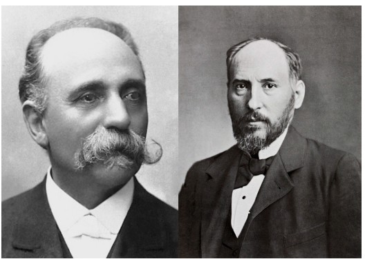
Both Golgi and Cajal were awarded the shared Nobel Prize in Physiology or Medicine in 1906 for their accomplishments in helping to understand “the structure of the nervous system.” Even though Cajal’s neuron doctrine was adopted widely by scientists, the elucidation of this organization would not have been made possible without Golgi’s development of the silver stain. The sharing of this prestigious award was ironic because of the many disagreements between the two scientists. Cajal commented on their relationship, saying:
“What a cruel irony of fate of pair, like Siamese twins united by the shoulders, scientific adversaries of such contrasting character!”
Cajal’s neuron doctrine was eventually given more support with the aid of modern techniques, like electron microscopy, that are capable of physically seeing the distance between two neurons. The neuron doctrine represents our current understanding of how the nervous system is organized, and this chapter is focused on describing the anatomical features of the nervous system at the cellular level.
Characteristics of neurons
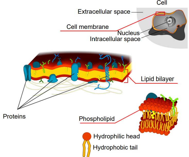
The main units of the nervous system are cells called . Although neurons do have a variety of adaptations that make them unique from other types of cells in the body, they are still cells. Therefore, they contain all of the basic features of a typical mammalian cell. For example, they are made up of an aqueous cytoplasm bounded by a cell membrane. This cell membrane, also called a plasma membrane or lipid membrane, consists of a sheet of several individual molecules called phospholipids, which consist of two hydrophobic (water-fearing) tails and a hydrophilic (water-loving) end. These phospholipids arrange themselves into a bilayer with the hydrophilic tails touching each other and the hydrophilic sides facing the cytoplasm and the extracellular space, which are both mostly water. Because of the chemical properties of the cell membrane, it is very effective at keeping ions and charged molecules separated, while allowing small molecules like water and oxygen across the cell.
They also have all the organelles that you would see in other cell types, like a nucleus and mitochondria.
The number of neurons in the adult human brain, according to our current best estimate, is close to 86 billion. This number was calculated using a revolutionary technique, the isotropic fractionator or “brain soup” developed by Brazilian neuroanatomist Suzana Herculano-Houzel. To put this number in context, we have about 37 trillion cells in the whole body, so neurons in the brain make up about 0.2% of all cells in the body.
Below are some unique characteristics that neurons have in common.
Neurons are electroactive
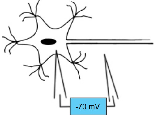
The inside of most cells have a negative electrical charge compared to the solution outside of the cell. This difference between the electrical charges is called the , or . When a neuron is at rest, the amplitude of this charge may be somewhere between -50 millivolts (mV) and -90 mV. Often times, we say that in general the cell rests at around -70 mV. Membrane potential is abbreviated as Vm.
Most neurons do not spend their entire lives at -70 mV. Instead, neurons have special proteins embedded in their cell membranes that allow for charged ions, such as sodium or chloride, to move into or out of the cell. When a net positive charge enters into the cell, the Vm becomes more positive. Likewise, when net negative charge enters the cell, the membrane potential becomes more negative. The membrane potential of a neuron can go from -70 mV to +45 mV and back to -70 mV in as quickly as two milliseconds!
Neurons are not the only electroactive cells in the body. Our lives depend on cells in the heart that are also capable of changing in potential, as they respond to rhythmic electrical impulses that drive our heartbeat. This is why being struck by lightning may cause your heart to desynchronize in activity, and why a shock from a defibrillator can electrically “kick-start” the heart back to a meaningful pattern.
Neurons are specialized for rapid communication
Many cells are capable of sending and receiving chemical signals across long distances and time scales. But neurons are able to communicate with a combination of electrical and chemical signals in a matter of milliseconds. Additionally, the shape of neurons and the organization of the neurons on a microscopic level make them effective for sending signals in a very specific direction. Many neurons have an incoming receiving end and an outgoing sending end. The placement of one neuron next to the correct partner is very important, and many chemical signaling systems are in place to ensure that the developing nervous system is properly wired together.
Neurons are “forever” cells
We are constantly replacing non neuronal cells. For example, the cells in our bones replace themselves frequently at a rate of about 10% each year. Our body makes new skin cells to replace the dying skin cells on the surface so that we have a “new” skin every month. The cells along the inside of our stomachs, exposed to very harsh acidic conditions, get replaced about every week. About 100 million new red blood cells are created every minute!

On the other hand, the mature nervous system generally does not undergo much , the creation of new neurons. The neurons that we have after development are the ones that we will keep until we die, and this permanence of neuronal count makes them different from almost every other cell of the body. However, the idea of adult neurogenesis is a topic of debate among neuroscientists, since some areas like the olfactory system and the hippocampus display new nerve cell production.
…But, neurons can change
Even though new neurons are not created in most areas of the brain, neurons still have the capability to change in their structure and function. Some of these changes, such as physical changes to the structures of the input sites of the neurons, are believed to last for a lifetime. We use the word to describe the ability for the brain to alter its morphology (derived from Greek plastikos, meaning “capable of being shaped or molded” – think of plastic surgery, where a person changes their physical appearance; see chapter 13, Leaning and Memory).
Also, neurons do have the capacity to repair themselves to some extent. Neurons of the PNS may get injured or completely destroyed as a result of trauma to the body. Afterwards, those injured neurons can regrow to connect once again with their original partner. This regrowth seems to depend on a few chemical signals that the body produces, such as nerve growth factor and brain derived neurotrophic factor. However, this process is often very slow, and does not always successfully restore the nervous system to the way it was pre-injury.
Cellular anatomy of neurons
The main function of neurons is to use changes in electrical properties in order to communicate with connected cells. This communication usually moves in one direction, and we will use this pathway as an outline for discussing the anatomical structures of the neurons.
Dendrites

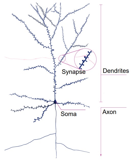
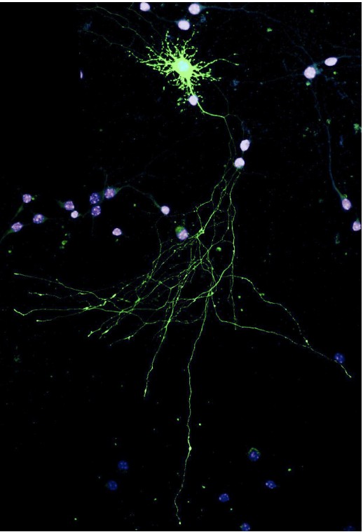
Information first enters the neuron through the , the branch-like extensions that protrude from the cell body. The word dendrite originates from the ancient Greek word “dendro-”, meaning “tree” (think of rhododendron). Dendrites look like the branches of a tree: they reach outward away from the center of the cell body, generally getting thinner the farther away you look.
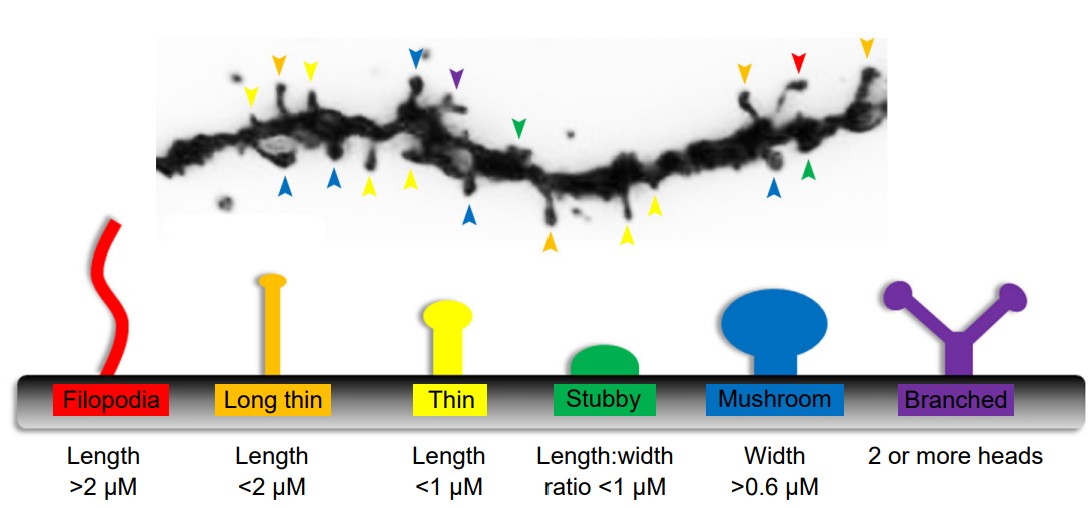
Along the dendrites of some neurons, it is possible to see tiny protrusions of cell membrane that stick out from the main dendrite. These bumps are called . Spines can roughly be classified based on their approximate shape; thin, mushroom, and stubby being some of the more easily recognizable forms. A spine may be about 100 nm in diameter, making it smaller than the wavelength of visible light, with a total volume of about 0.1 femtoliter – one ten-quadrillionth of a liter!
Chemical signals released by another cell are received by the dendritic spines, and so each spine may represent an input site of communication. Some cells, like a pyramidal neuron in the hippocampus, may have more than 30,000 spines, indicating that one cell may detect information from several incoming cells.
We believe that spines are one of the most important sites where the nervous system is able to change. For example, neurons change shape after exposure to various environmental conditions, such as stress or exposure to drugs. Tiny changes to the surface of the neuron at the level of dendritic spines is an example of plasticity. Dendritic plasticity is thought to underlie the reason that we can learn new facts or maintain memories about our childhood over long periods of time. Some set of tiny, submicroscopic changes to the morphology of dendritic spines may represent a single complex memory that you form.
A neuron does not need spines for receiving information or for plasticity to take place. Many cells lack spines, but are still capable of permanently changing. The input site may be anywhere along the dendrite, or even at the cell body, the “center” of the neuron.
Cell body (soma)
Information that arrives through the many dendrites of a neuron eventually filters into the , or the , of the neuron. Somata (plural of soma) vary in size across different types of neurons, the largest somata belonging to the Betz cells of the motor cortex with an area upwards of 100 microns in diameter: about the size of a single grain of salt. On the opposite end of the spectrum, granule cells of the cerebellum are so densely packed so that they make up more 70% of all neurons in the brain, maybe 4 microns in diameter.
The cell body contains many of the organelles that are essential for the production of proteins that the neuron needs. Many of these organelles are not unique to neurons, and can be found in other cell types. The most apparent organelle visible under high magnification (400x) is the , which houses DNA and other genetic material. From our understanding of the central dogma of molecular biology, this DNA is transcribed into a string of single-stranded genetic code called (mRNA), which is exported out of the nucleus. This mRNA is then used as a guide for the synthesis of proteins.
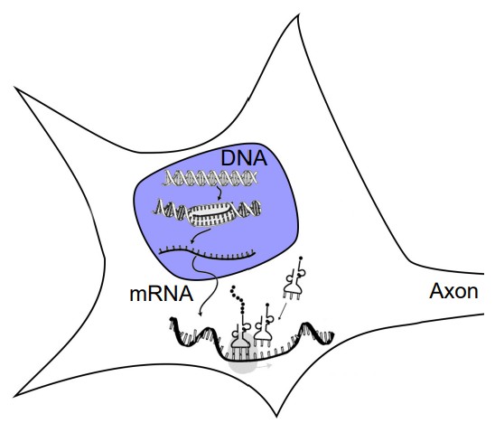
The next step of protein creation depends on organelles that are adjacent the nucleus. Physically continuous with the membrane that surrounds the nucleus is a folded membranous organelle called the (ER). Attached to the ER are several , which are the molecular machines that read the mRNA and translate that code into proteins. The are layers of folded plasma membranes that function in transport. They are found near the nucleus, although small protrusions of these organelles may reach into the other parts of the neuron.
Axon
The is the main output extension of the neuron. While neurons only have a single axon extending from the cell body, this axon can branch several times after exiting the soma. In branching, an axon from a single neuron is able to communicate with many other neurons at the same time. Axons are usually thinner than dendrites, some being only a micron in diameter. Several axons can bundle and travel together; these are . Axons can be very long; the longest axon in the human body is part of the sciatic nerve that runs from the posterior end of the spinal cord down the leg to control the muscles of the big toe.
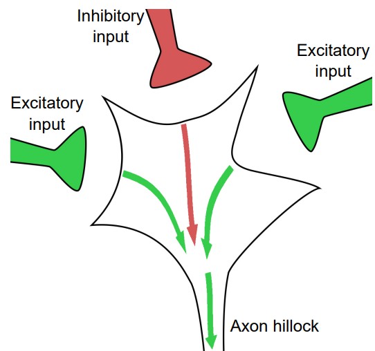
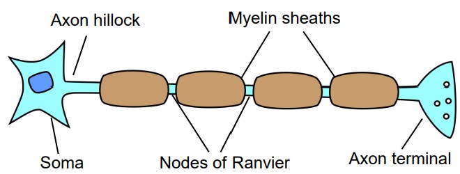
The first section of the axon, the junction between the cell body and the beginning of the projection, is a patch of axonal membrane with very unique characteristics. This section is called the . With respect to signal transduction, the axon hillock works as the integrative center of the neuron. It is responsible for deciding whether to pass a signal onto the next cell. The cell membrane of the hillock performs a complex set of “cellular arithmetic” that weighs all of the incoming signals: excitatory, inhibitory, and modulatory signals. After all of the calculations have been performed, the membrane either sends a signal or not.
At the end of each branch of the axon is a small swelling, which is called the or . The terminal is the part of the neuron that is specialized for the production and release of the neurotransmitters that are used for communication between neurons. One subarea of specific interest in the axon terminal is a small patch of membrane called the . Embedded in the cell membrane at the active zone is a variety of proteins that are important for the process of neurotransmitter release.
Inside the axon
Most of the proteins synthesized in neurons are created in the cell body very close to the nucleus, where the mRNA that is exported from the nucleus is able to easily interact with the rough endoplasmic reticulum and the ribosomes. But, some of these proteins are needed far away from the cell soma, at the axon terminal, for example. Neurons need some system of transport system that can move newly created proteins to where they need to go. Inside the length of the axon runs an organelle called , which function like a molecular railway for proteins. The cells use motor-like proteins that can carry other proteins along the microtubules. When substances are transported away from the cell body, it is called anterograde transport, while the process of moving proteins towards the cell body is retrograde transport.
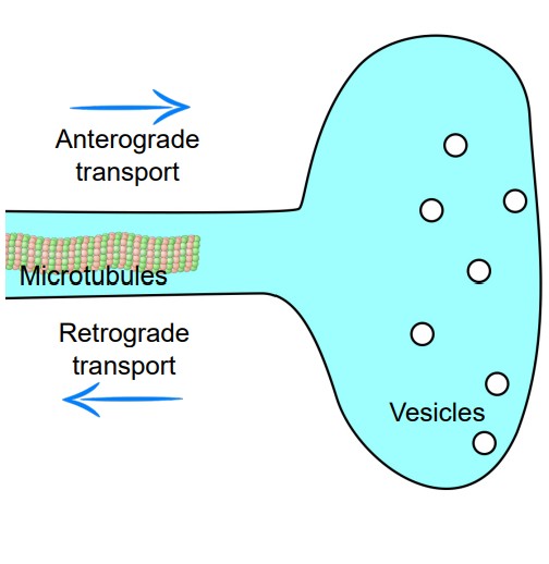
Another important organelle that is found along the length of the axon are . These organelles are made up of several different proteins that serve as a cellular “scaffolding” that helps keep the structure of the axon intact. Mature neurons can be very dense with neurofilaments, which increases the diameter of the axon.
Within the terminal are a number of , small spherical “packages” made of cell membrane that are coated in special proteins. Within these vesicles are the molecules that the neurons use for chemical communication. When the action potential travels down the axon and reaches the axon terminal, the cell membrane changes in electric charge, and this causes vesicles to fuse with the inner membrane of the neuron. During fusion, the vesicular membrane merges with the cell membrane at the axon terminal, causing the contents of the vesicle to be released outside the cell.
Outside the axon
Some neurons have a special modification surrounding their axon called a . Myelin is comprised of several tightly-wrapped layers of cell membrane that encompasses a short section of the axon. Myelin might be wrapped as many as 250 or 300 times around a single section of axon. Myelin does not extend fully enclose the entire length of the axon from soma to terminal, but rather surrounds short sections at a time. The spaces of exposed axon between each section of myelin are called . On average, these nodes are about 1 micron long.
Myelin serves a few functional purposes. Myelin increases the speed by which an electrical signal is transmitted. Some of the most heavily myelinated axons are able to send signals up to 120 meters per second (almost 270 miles per hour, faster than a Formula 1 racer.) Myelin also increases the effective thickness of the cell membrane along the axon. In doing so, myelin acts as an insulator that causes signals to more reliably be passed down the axon.
Synapse
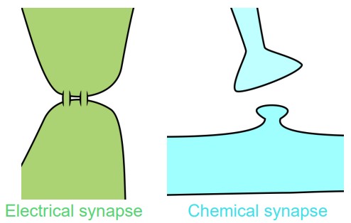
The synapse is the physical distance that separates two neurons. In agreement with Cajal’s neuron doctrine, the nervous system is not made up of a single cell with a giant, shared cytoplasm, but rather a series of neurons in close proximity, separated by a gap of extracellular space.
This distance between two cells can vary depending on the nature of the synapse. An electrical synapse may be less than 5 nanometers apart. Cells connected by electrical synapses share cytoplasm but have two separate cell membranes. On the other hand, a chemical synapse is a larger distance, about 15 – 40 nm Figure 3.14 Electrical synapses physically share cytoplasm (left) while chemical synapses use neurotransmitters to communicate (right). Electrical synapse Chemical synapse across. Adjacent neurons connected by chemical synapses do not share cytoplasm
Clinical connection: Multiple sclerosis
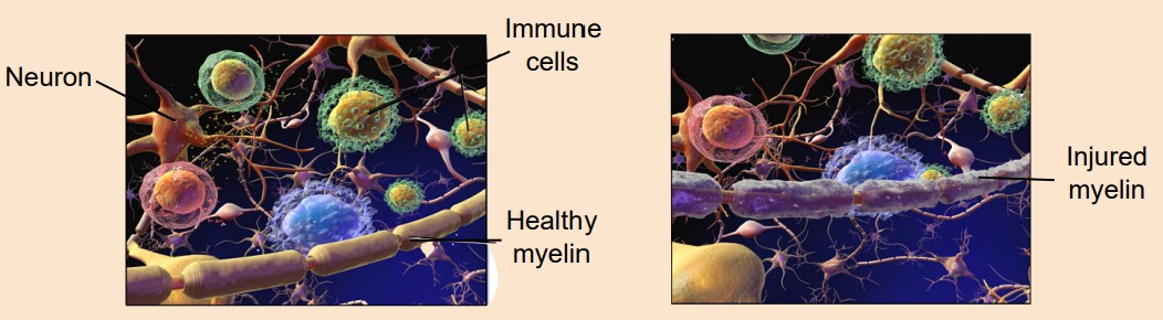
(MS) is a disease that results from destruction of myelin in the CNS. When myelin is damaged, signals do not reliably propagate from the brain to the body, from the body to the brain, or between areas of the brain. MS damages myelin at both descending and ascending neurons, so a person with MS might experience muscle weakness, poor balance, and muscle spasms (efferent motor neurons) as well as numbness and pain (afferent motor neurons). It can also affect neuronal signaling in the brain, causing symptoms such as cognitive impairment, vision loss, and changes in affect leading to depression. Multiple sclerosis is a common neurological disorder that affects some 2.5 million people worldwide. MS typically presents in adulthood, usually between 20- 50 years old. Unfortunately, there is currently no known cure for MS. Therapy is focused on either slowing the progression of the disease, helping patients recover after an attack, or decreasing the severity of the symptoms. While MS has the potential to be debilitating and painful, the disease itself is not necessarily lethal, and MS only decreases life expectancy by 5-10 years on average. People with MS are just as likely to die from natural causes as much as a neurotypical person. One of the leading theories about the cause of MS is a faulty immune system. A normally functioning immune system identifies and destroys foreign pathogens. Sometimes, this system makes a mistake, and the immune response recognizes normal parts of the body as being a “foreign object”, causing the body to destroy itself. This is called an autoimmune disorder. In the case of MS, it is believed that the immune system identifies and targets myelin for destruction, leading to demyelination. Although immunosuppressants may slow the progression of MS, these drugs increase the risk of a person developing an illness that a healthy immune system would be able to prevent.
Classifications of neurons
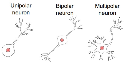
Neurons have a wide variety of shapes and can be divided roughly into three different classes depending on their morphology. In order of increasing morphological complexity, they are:
1. Unipolar cell
Unipolar cells have a single cellular extension coming off the soma that acts as both the receiving and the sending end. Unipolar cells are very common among invertebrates; humans do not have unipolar neurons.
2. Bipolar cell
A bipolar cell, as can be implied from the name, has a single dendrite and a single axon. They are not very common, but can be found in human sensory systems, such as in the eye or in the signaling pathway connecting the ear to the brain.
3. Multipolar cell
Multipolar neurons have several dendrites and one single axon. These cells are the most common among all the neurons in the human nervous system. Most drawings of neurons you will see in this book are multipolar cells.
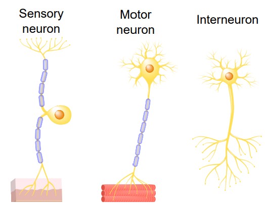
In addition to classifying neurons based on their morphology, neurons can also be divided based on their functions.
1. Sensory
Sensory neurons are the afferent neurons that are responsible for obtaining information about either the outside world or the internal environment and passing that information towards the CNS. Sensory neurons can detect a variety of stimuli, ranging from photons of light (visual system) or chemicals floating in the air (olfactory system), to carbon dioxide levels in the blood (chemoreceptors) or stretching of the muscles (spindle receptors). Since they are highly specialized for detecting different types of stimuli, sensory neurons have a large variety of shapes and structures.
2. Motor
Motor neurons, or motoneurons, carry signals from the CNS to the body. There are two main types of motor neurons. Somatic motor neurons control skeletal muscle movement, such as flexing or extending the muscles of the arm. They release neurotransmitter directly onto muscles. Autonomic motor neurons, on the other hand, release neurotransmitter onto a clump of neurons outside of the CNS called the autonomic ganglion, which then signal to the smooth muscle, cardiac muscles, or glands. 3. Interneuron Interneurons exist as a relay between the sensory or motor neurons and the CNS, or between each other. They represent a very broad class of neurons, and make up an important part of reflex circuits, such as the knee-jerk response.
The above classifications are only a rough guideline for separating neurons. As with most biologists, neuroscientists enjoy classifying cells based on their properties. It is almost always preferable to use the most specific name possible in identifying neurons. Many cells have been identified based on the way they look (chandelier cells), some named on the neurotransmitter released (cholinergic interneurons), some named by where they are found (cerebellar granule cell), and yet others named based on the person who discovered them (Purkinje cells)
Visualizing the synapse
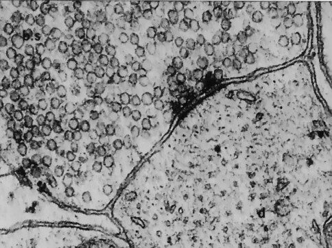
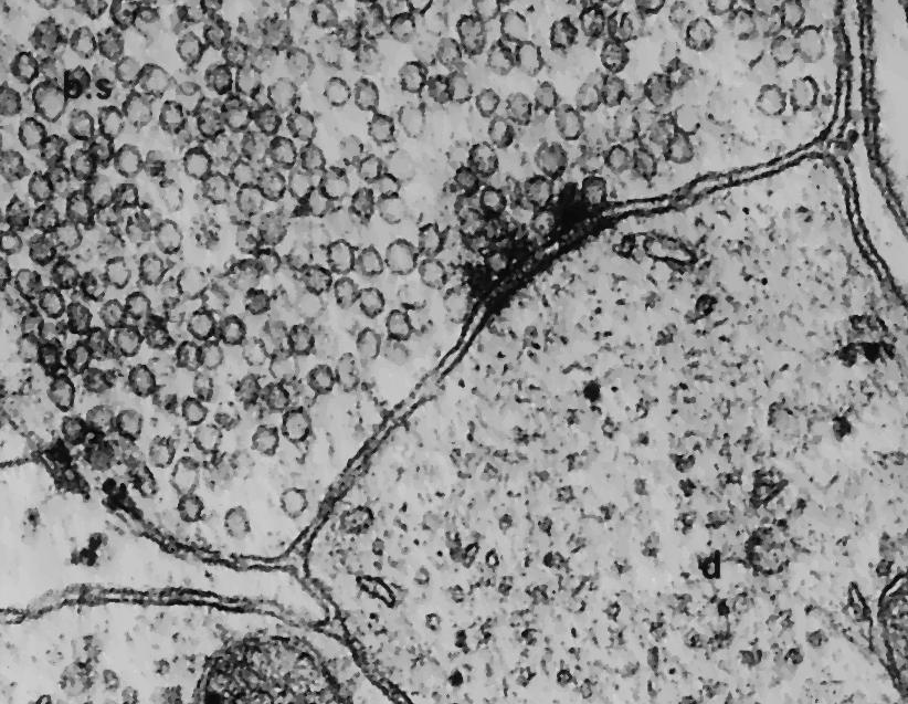
The original microscopes that van Leeuwenhoek used to visualize cells and microorganisms relied on the transmission of light to see objects up close. For us to be able to see objects, light needs to bounce off the intended target. Light particles travel in a wave, and if the object we intend to visualize is smaller than the wave, then the wave would pass right over the object. The shortest wavelength of visible light is around 380 nanometers, so light microscopy is limited to visualizing objects in the micrometer or larger range – a 2,000x magnification with the best possible lenses. In the early 1950s, a group of scientists developed a technique called (EM). In EM, a beam of electrons is aimed at a sample in a vacuum, and the reflection of those electrons can be collected and detected with a computer. Since electrons have a much shorter wavelength than visible light, you are able to resolve objects as small as 50 picometers – more than a 10,000,000x magnification! Using EM, you are able to get ultrastructural resolution of the synapse. In two adjacent neurons, EM allows you to distinguish the boundaries of both neurons, and to clearly see the synapse in between. You can also see the vesicles contained in the axon terminal, sometimes in the middle of fusion.
Cellular functions of glia
Although most of neuroscience is concerned with understanding the functions of neurons, there are other cells in the nervous system that are just as interesting. These cells are grouped together under the umbrella classification of . Historically, when these non-neuronal cells were visualized under the microscope, the histologists and anatomists had no idea about their function. They were seen all around the neurons, so the assumption was that these cells were structural elements, a sort of living glue that held the nervous together. Today, we know that these glia serve all variety of functions; unfortunately the misnomer “glia,” derived from the Latin word for glue, is still used to describe these non-neuronal components of the nervous system. We estimate that the brain has roughly an equal number of glial cells and neurons—86 billion of each.
There are many different classes of glia, but we will focus on five types.
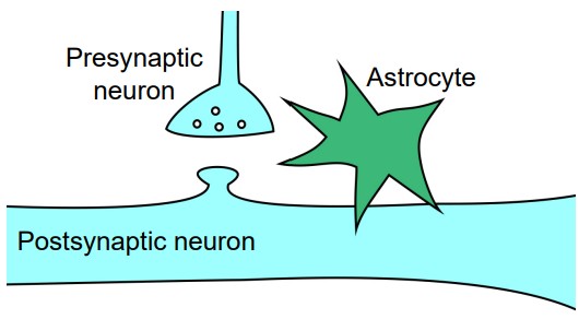
1. Astrocytes
Astrocytes are named for their characteristic star-shaped morphology. Astrocytes have a dense expression of the protein (GFAP), and this protein is often used as a marker for differentiating astrocytes from other cell populations.
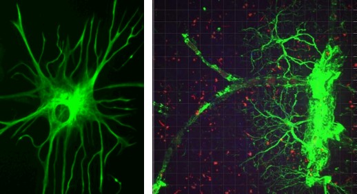
One of the main functions of astrocytes in the brain is to help maintain the blood-brain barrier. At the end of the extensions of the astrocyte are protrusions called “endfeet.” These endfeet are often wrapped around the endothelial cells that surround the blood vessels. The endfeet release important biological compounds that allow the endothelial cells to remain healthy as they function in maintaining the blood-brain barrier.
Astrocytes are also very closely associated with synapses. Astrocytes have a very dense expression of proteins on their cell surface that can transport molecules of the neurotransmitter glutamate, for example, inside the astrocyte. By acting as a glutamate “sponge,” astrocytes are able to decrease the strength of a glutamate signal. Through a similar uptake mechanism, astrocytes can also affect the extracellular concentration of ions such as potassium, which then has an influence on cellular excitability. Because of these interactions between astrocytes and neurotransmission at the synapse, we use the phrase to refer to the three components of a synapse: The presynaptic neuron, the postsynaptic neuron, and the astrocyte.
Astrocytes also synthesize and produce a variety of , which are helper molecular signals that serve several different functions. For one, trophic factors signal to neurons that the neuron should continue to live, or that specific synapses should be maintained. They help guide the neurons as they reach out, forming synapses where appropriate.
2. Oligodendrocytes
Oligodendrocytes only exist in the CNS. Their name is derived from their morphology. Oligo- refers to “a small number” (think oligarchy, a government ruled by a few people), and dendro- refers to “tree” (like a rhododendron). Each oligodendrocyte has a few branches that reach away from the cell body.
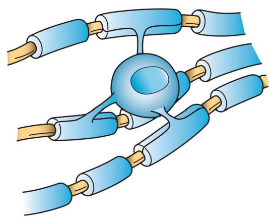
The main function of the oligodendrocytes is to add a layer of myelin around the axons of nearby CNS neurons. A single oligodendrocyte is able to myelinate up to 50 segments of axons. As cells that produce myelin, they are responsible for increasing the conduction speed of nearby neurons as they send signals. When the oligodendrocytes begin myelinating, they are able to produce almost 3 times their weight in membrane per day. By maturity, the oligodendrocyte supports cell membrane that is 100 times the weight of the cell. Due to the need to support such large amounts of myelin, it’s estimated that they have the highest metabolic rate of any type of cell in the brain.
3. Schwann cells
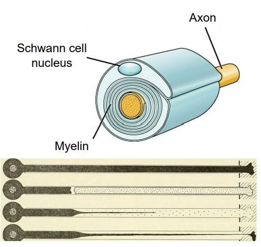
Schwann cells are named after the German biologist who first described these glial cells. He found that there are cells that are wrapped around the axons of nerve cells that project towards muscles – myelin. These Schwann cells can only be found in the PNS.
The main action of Schwann cells is to provide a section of myelin sheath for PNS neurons, and in this way, they function similarly to the oligodendrocytes. Schwann cells produce only a single section of myelin, compared to oligodendrocytes, which myelinate multiple sections.
Schwann cells also function in the regeneration of injured axons. When nerves in the PNS are damaged after trauma, Schwann cells rapidly mobilize to the site of injury. Here, the remaining loose myelin hinders the regeneration process, and the Schwann cells destroy the extra cellular membranes. Schwann cells also produce signaling molecules that guide the injured axons to the correct targets, which helps the axon regrow.
4. Microglia
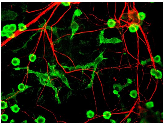
Microglia are a bit different from the other glial cell populations. For one, microglia are more immune cells rather than neural. They act as cellular scavengers that travel throughout the brain and spinal cord. It is estimated that microglia make up 10-15% of all cells in the brain.
As immune cells, microglia identify and destroy clumps of proteins, dead / dying cells, or foreign pathogens that enter into the brain. After an injury to the CNS, like a traumatic blow to the head, microglia rapidly react to the area of the insult. The marker Iba1 is often used to identify when microglia are reacting to an injury.
5. Ependymal cells
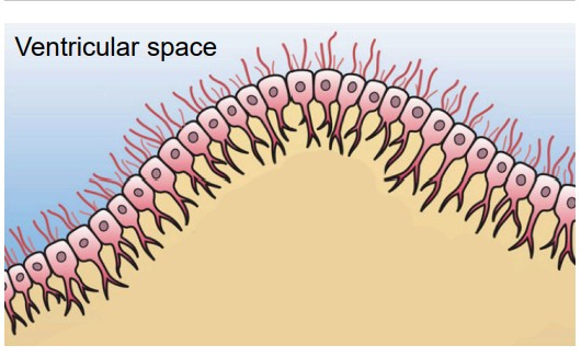
ventricles and produce cerebrospinal fluid.
Along the inside of the ventricles are a lining of glia called the ependymal cells. These ependymal cells are columnar with small finger-like extensions called cilia that extend into the ventricles and into the central canal that runs down the inside of the spinal cord.
Ependymal cells produce CSF. In total, the body can make about half a liter of CSF each day (a little more than two cups.) The ependymal cells are part of a structure called the , the network of blood vessels and cells that form a boundary between the blood and the CSF.
Image Credits
Figure 1 .1: https://commons.wikimedia.org/wiki/File:Figure_35_03_05_Brain_size_Vertical.png
Figure 1.2: https://commons.wikimedia.org/wiki/File:Action_potential_basic_shape.svg Modified by Austin Lim https://commons. wikimedia.org/wiki/File:Acetylcholine.svg https://commons.wikimedia.org/wiki/File:Resting_Potential.png Modified by Austin Lim https://commons.wikimedia.org/wiki/File:Glutamate_2,3-aminomutase_general_reaction.png Modified by Austin Lim
Figure 1.3: https://commons.wikimedia.org/wiki/File:Brain_image_of_blind_echolocator.tif Modified by Austin Lim
Figure 1.4: https://commons.wikimedia.org/wiki/File:The_Scientific_Method.svg Modified by Austin Lim
Figure 1.5:
Figure 1.6: https://commons.wikimedia.org/wiki/File:Adverse-childhood-experiences-study-psychological-trauma-childhood- trauma-strengthen-prevention-thumbnail.jpg Modified by Austin Lim
Figure 1.7: https://commons.wikimedia.org/wiki/File:Phineas_Gage_CGI.jpghttps://commons.wikimedia.org/wiki/ File:Phineas_Gage.jpg
Figure 1.8: https://pixabay.com/photos/light-red-stop-street-427961/
Figure 1.9: https://commons.wikimedia.org/wiki/File:Brain_weight_age.gif Modified by Austin Lim
Figure 1.10: https://en.wikipedia.org/wiki/Contralateral_brain#/media/File:AxialTwistDevelopment.png
Figure 1.11: https://commons.wikimedia.org/wiki/File:WLANL_-_andrevanb_-_kist_uit_de_27-_31e_dynastie_(4).jpg https:// commons.wikimedia.org/wiki/File:Three_of_four_cranial_crochets,_copies,_used_for_removing_th_Wellcome_L0058415. jpg
Figure 1.12: https://commons.wikimedia.org/wiki/File:Paul_Broca_2.jpg https://commons.wikimedia.org/wiki/File:Pierre_Marie,_ Travaux_et_memoires._Wellcome_L0028667.jpg
Figure 1.13: https://commons.wikimedia.org/wiki/File:Scanning_electron_microscope_-_UFCH_JH_(2020)_01.jpghttps:// commons.wikimedia.org/wiki/File:Synapse_neuro-neuronale.png
Figure 1.14: https://commons.wikimedia.org/wiki/File:Pyramidal_hippocampal_neuron_with_fake_microelectrode.jpg https:// pixabay.com/illustrations/psychology-mind-thoughts-thought-2422442/ https://commons.wikimedia.org/wiki/File:Caffeine_ and_adenosine.png https://commons.wikimedia.org/wiki/File:ActionPotential.png https://commons.wikimedia.org/wiki/ File:Brain_network.pnghttps://upload.wikimedia.org/wikipedia/commons/4/40/Deep_Brain_Stimulation_surgery.png https://commons.wikimedia.org/wiki/File:Human_brain_NIH.png?wprov=srpw1_219
3.1 https://upload.wikimedia.org/wikipedia/commons/6/6d/GolgiStainedPyramidalCell.jpg
https://upload.wikimedia.org/wikipedia/commons/1/1a/Human_hippocampus_2.5x.jpg
3.2 https://upload.wikimedia.org/wikipedia/commons/5/57/Camillo_Golgi_nobel.jpg
https://upload.wikimedia.org/wikipedia/commons/3/30/Cajal-Restored.jpg
3.3 https://upload.wikimedia.org/wikipedia/commons/5/5f/Cell_membrane_detailed_diagram_
simplified_ca.svg modified by Austin Lim
3.4 https://svgsilh.com/9e9e9e/image/2040692.html modified by Austin Lim
3.5 https://pixabay.com/photos/axe-old-lumberjack-blade-1008981/
3.6 https://upload.wikimedia.org/wikipedia/commons/0/0a/Anatomy_of_a_Neuron_with_Synapse.png
modified by Austin Lim
3.7 https://upload.wikimedia.org/wikipedia/commons/7/74/Dendritic_arborization_patterns_2.png
https://upload.wikimedia.org/wikipedia/commons/9/90/Submerged_Albizia_Saman_in_the_Mekong_
at_sunset_%28close-up_view%29.jpg
3.8 https://upload.wikimedia.org/wikipedia/commons/6/6e/Geometric_characteristics_of_dendritic_
spines.png modified by Austin Lim
3.9 https://upload.wikimedia.org/wikipedia/commons/f/fb/MRNA-interaction.png modified by Austin
Lim
3.11 Image courtesy of Connor Maltby
3.12 https://upload.wikimedia.org/wikipedia/commons/8/86/201704_microtubule.svg modified by
Austin Lim
3.15 https://upload.wikimedia.org/wikipedia/commons/2/2a/Multiple_Sclerosis.png modified by Austin
Lim
3.16 https://upload.wikimedia.org/wikipedia/commons/9/92/Neurons_uni_bi_multi_pseudouni.svg
modified by Austin Lim
3.17 https://upload.wikimedia.org/wikipedia/commons/6/6c/Synapse_neuro-neuronale.png
3.19 https://upload.wikimedia.org/wikipedia/commons/5/56/Human_astrocyte.png
https://upload.wikimedia.org/wikipedia/commons/3/34/Astrocytes_Wrapping_around_Blood_Vessel.
png
3.21 https://upload.wikimedia.org/wikipedia/commons/2/2e/Oligodendrocyte_illustration.png
3.22 https://upload.wikimedia.org/wikipedia/commons/5/52/CeluladeSchwann.jpg
https://upload.wikimedia.org/wikipedia/commons/0/03/Analysis_of_
development_%281955%29_%2817549733973%29.jpg modified by Austin Lim
3.23 https://upload.wikimedia.org/wikipedia/commons/0/0b/Microglia_and_neurons.jpg
3.24 https://upload.wikimedia.org/wikipedia/commons/c/ca/Glial_Cell_Types.png modifed by Austin
Lim
The main active cellular units of the nervous system.
The electrical output of many neurons is an all-or-nothing response called an action potential.
A wide variety of chemicals that are responsible for passing information between neurons.
In an experiment, the scientist uses a stepwise process of developing a research question and hypothesis, then answering that question by performing tests.
One factor that is being changed.
The factor that is influenced.
Extraneous variables.
A group that is as similar as possible in every way except for the manipulation of the independent variable.
A change in the independent variable causes a change in the dependent variable.
The distribution of the characteristics in the sample is proportionally similar to the distribution in the total population
A type of observational study.
A highly detailed description of a single patient and their condition.
The sites of close contact between neurons.
The process in which new neuronal growth occurs in the brain.
the ability to change over time
Tnere are some conditions that are neurodegenerative, meaning that their symptoms get progressively worse over time
Signaling between the brain and the rest of the body: signaling pathways from the left brain crosses over to communicate with the right half of the body, and vice versa.
In the mid-1800s, Paul Broca was one of the first to suggest that specific areas of the brain were responsible for carrying out specific functions, which came to be called localization theory.
Suggests that behavioral functions require activation of cells across several different areas of the brain.
the act of generating knowledge through a combination of senses, memories, and thoughts
A type of microscope that uses electrons to produce images.
A method to render an entire brain transparent, which helps us to map out the nature of the connections that span the nervous system.
The discussion that centered around the question if a function as complex as consciousness could result from activity of a clump of cells.
The idea that the whole is greater than the sum of its parts.
originating from within the body
they originate from outside the body
the use of mathematical modeling to describe or predict some aspect of the nervous system.
Neurons that were stained black resulting from a technique using a silver compound that caused the silver to precipitate inside the cell membranes.
the idea that the parts of the nervous system are all one very large, physically connected network
the nervous system is a series of individual units that are separated from one another physically
The difference between the electrical charges inside and outside the cell
The difference between the electrical charges inside and outside the cell
the branch-like extensions that protrude from the cell body
tiny protrusions of cell membrane that stick out from the main dendrite
Receives information from dendrites of a neuron
another term for a cell body
houses DNA and other genetic material
DNA is transcribed into a string of single-stranded genetic code called messenger RNA
the folded membranous organelle that surrounds the nucleus
molecular machines that read the mRNA and translate that code into proteins
layers of folded plasma membranes that function in transport
main output extension of the neuron
Several axons bundled together
a patch of axonal membrane with very unique characteristics
Small swelling at the end of each branch of the axon
another term for the axon terminal
A subarea in the axon terminal containing a small patch of membrane that are important for neurotransmitter release
functions like a molecular railway for proteins
made up of several different proteins that serve as a cellular “scaffolding” that helps keep the structure of the axon intact
small spherical “packages” made of cell membrane that are coated in special proteins
comprised of several tightly-wrapped layers of cell membrane that encompasses a short section of the axon
The spaces of exposed axon between each section of myelin
a disease that results from destruction of myelin in the CNS. People with MS might experience muscle weakness, poor balance, muscle spasms, numbness and pain, and weakened neuronal signaling in the brain
a technique using a beam of electrons aimed at a sample in a vacuum, and the reflection of those electrons can be collected and detected with a computer
non-neuronal cells in the nervous system and brain
often used as a marker for differentiating astrocytes from other cell populations.
refers to the three components of a synapse: The presynaptic neuron, the postsynaptic neuron, and the astrocyte.
helper molecular signals that serve several different functions
the network of blood vessels and cells that form a boundary between the blood and the CSF
