
Emotions, Aggression, and Stress
Additional Videos and Readings
Video: Author/Source: Ted Talks; How stress affects your brain – Madhumita Murgia
Chronic stress can have significant negative effects on the brain, including changes in its structure and function. It is triggered by the release of cortisol, which can lead to increased activity in the amygdala and decreased function in the hippocampus. Chronic stress can also shrink the brain and make it harder to learn, remember, and may contribute to mental health problems. The effects of stress can be inherited through epigenetic changes. However, exercise and meditation are effective ways to counter these effects, reducing stress and improving brain health.
Video: Author/Source: Ted Talks; The biology of our best and worst selves – Robert Sapolsky
In his TED talk, Robert Sapolsky discusses the complex nature of biology, environment, and behavior and their role in shaping human behavior. He touches on the role of genes, hormones, and the brain in influencing our actions, both at our best and worst moments. Sapolsky emphasizes that our behavior is not solely determined by nature or nurture but rather a dynamic interaction between the two. He also highlights the potential for positive change by understanding the biological basis of our actions and the possibility of empathy and compassion.
Reading – Harrell et al., 2011 – Du Bois Review – Multiple pathways linking racism to health outcomes
At the airport, you can observe people experiencing a wide range of emotions: Sadness at seeing family members off, fear and anxiety for those about to fly for the first time, love when a long-distance relationship is reunited, and anger over unpredictable cancellations.
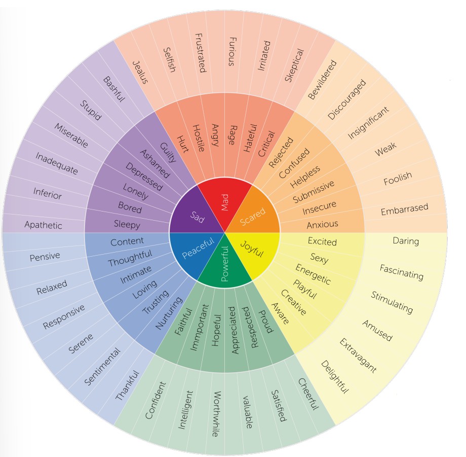
Emotions are complex neurophysiological states that contribute to an internal feeling and guide behavior. Some emotions are pleasant (joy), some are negative (disgust), and some are a mix of both (nostalgia). Some are short-lasting (surprise) while others may persist over years (vengefulness).
However, beyond this statement, it becomes very difficult to put a clear-cut definition on “emotion”. The difficulty with defining emotion arises because of the fluid nature of emotions: they exist on a spectrum, multiple emotions are experienced simultaneously, each emotion is perceived by different people in unique ways, and everyone has a slightly different interpretation and understanding of an emotion.
The field of seeks to understand the neural mechanisms that underlie emotion. The field has expanded with the help of functional imaging methods like EEG and fMRI, where changes in brain activity can be measured and quantified as a person experiences different emotion-provoking stimuli. Affective neuroscientists work to develop biology-supported therapies for disorders such as depression, PTSD, and addiction, which are dysregulations of the emotions of sadness, fear, and desire, respectively.
15.1 A History of Emotion Research
Origins of emotion
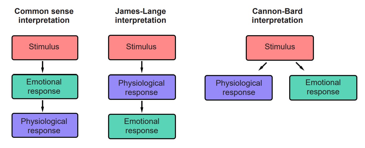
One of the older theories about the origin of emotion is based on the most common sense interpretation of cause and effect. For example, imagine some noticeable emotional stimulus, such as encountering a hungry lion on the sidewalk. The logical cause and effect explanation suggests that seeing the lion prompts the emotion of fear, which then causes the sympathetic nervous system “fight-or-flight” response (elevated heart rate and blood pressure, increased respiration, and cellular mobilization of energy).
In the 1880s, psychologist William James and physician Carl Lange independently developed a new theory about the origin of emotion. According to the , and contrary to a common sense understanding of the origin of emotion, the body’s physiological changes precede the onset of an emotional response. For example, imagine encountering that same hungry lion on the sidewalk. The James-Lange theory tells us that the perception of the threat of being eaten causes the sympathetic nervous system response, and that these physiological changes trigger the onset of fear.
Soon after, in the 1920s and 30s, two physiologists named Walter Cannon and his doctoral student Philip Bard criticized the James Lange theory. In one experiment, they surgically removed the entire sympathetic nervous system from cats, destroying the nerves that regulate vascular dilation, the activity of liver enzymes, and the reaction that causes the hair standing on end. These cats were then put before a threatening aggressor. If the James-Lange theory was true, then the physiological changes should precede the emotive response. However, the cats exhibited the fear / aggression response (such as posturing, hissing, and clawing) even without an intact sympathetic nervous system. Relatedly, patients with spinal cord injuries have a similar lack of autonomic inputs to the brain, but their capacity for emotional responding is still intact.

In a second criticism, Cannon and Bard proposed that the physiological changes seen in sympathetic nervous system activity may arise for a variety of reasons, not always for emotionally salient reasons. For example, intense exercise causes strong cardiorespiratory changes; however, we do not necessarily feel a strong emotional state after this physiological perturbation. Likewise, exogenous administration of epinephrine, onset of fever, or being in cold temperatures may also trigger some physiological changes without causing a strong emotional response.
Based on their evidence opposing the James-Lange theory, Cannon and Bard developed an alternative explanation for the origin of emotions. According to the Cannon-Bard theory of emotion, the perception of an emotionally charged stimulus prompts simultaneous but independent activation of both the autonomic nervous system and the emotional response.

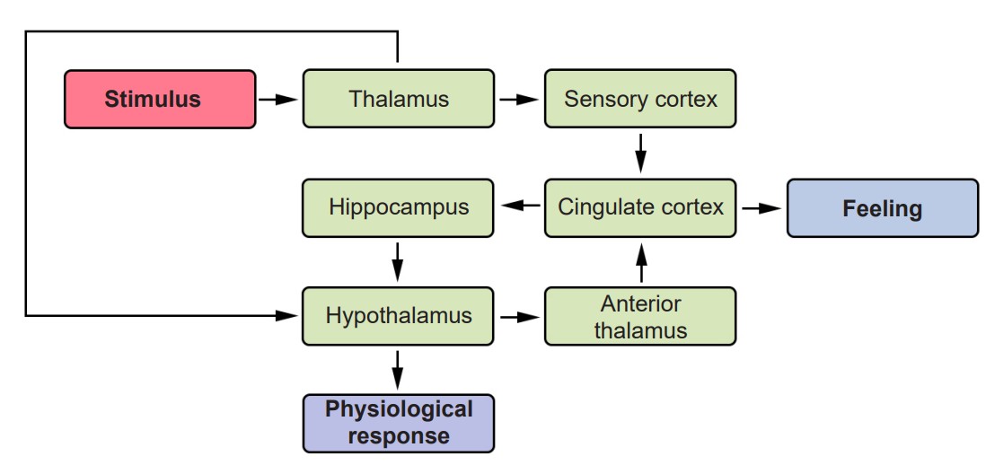
The Cannon-Bard theory also draws attention to the neuroanatomical structures that trigger the autonomic and emotional responses, with a particular focus on diencephalon structures such as and . To localize the anatomy behind emotional signaling, researchers performed a series of lesion experiments, systematically injuring different parts of the brain. To their surprise, when the cortex of the cat was surgically separated from the rest of the nervous system, a procedure called the , the cats would exhibit a hyper-aggressive response to stimuli. For example, an innocuous touch of the tail would trigger violent clawing, biting, and hissing, behaviors which were described as . However, when they lesioned the thalamus, sham rage was no longer observed. They concluded that rage (and other powerful emotions) is normally under inhibitory control by the cortex.
Just years later, in 1937, American neuroanatomist James Papez (pronounced payps) ascribed emotional behavior to a particular series of brain structures. These structures, collectively called the , consist of the hypothalamus, cingulate gyrus, thalamus, hippocampus, and more. Papez observed unusual aggression among animals with injury to these structures, suggesting that emotional responding is distributed across many areas, rather than localized. (Notably missing from the Papez circuit structures is the amygdala, which was later added in a future revision in the 1950’s.)
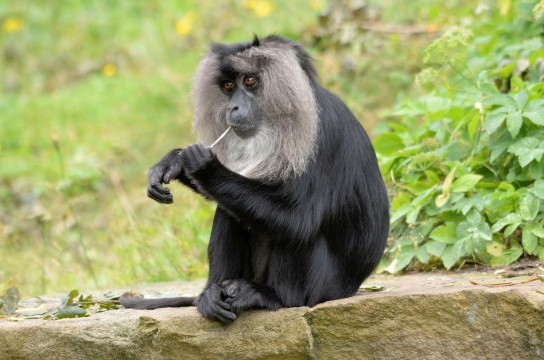
In 1939, two researchers named Heinrich Kluver and Paul Bucy described a unique set of emotional deficits in monkeys with bilateral temporal lobe removal, further providing evidence of the neuroanatomy of emotion. These animals fail to display fear or anger, even in the face of life-threatening stimuli such as a large snake. They also display (the inability to recognize faces or objects visually), psychic blindness, hypersexuality, and (an inappropriate fixation with using the mouth to interact with surroundings, such as licking or eating nonfoods). Collectively, these sets of symptoms are called . Although it is primarily an experimental manipulation observed in monkeys, some human patients may develop the condition, such in after brain surgery, stroke, viral encephalitis, traumatic brain injury, and many others.
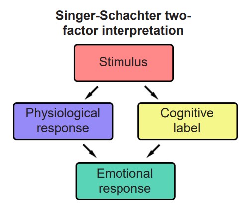
A new angle to the origin of emotion was proposed by Stanley Schachter and Jerome E. Singer in 1962. They were interested in how the same physiological response can be attached to two wildly different emotions – consider the rise in heart rate and respiration that are associated with encountering that lion (fear), or when you receive wonderful news (elation), or when you make eye contact with your romantic partner (love). According to their , people use a combination of the physiological response and a cognitive label to determine the emotion that is most appropriate for a given circumstance. The cognitive label comes from two parts. One is prior knowledge, such as the thought processes “lions eat meat if they are hungry, so I should be afraid.” The other part is observing environmental cues, such as “everyone around me is running away and screaming, so I should also be afraid.”
To test their theory, Schachter and Singer administered epinephrine to patients. Epinephrine is a hormone that produces the physiological signs very similar to those observed in a sympathetic nervous system response: elevation of heart rate, respiration, blood flow to muscles, and energy utilization. Then, the patient is put into a waiting room with another “patient” – in actuality an undercover member of the research team (the confederate). For one set of patients, the confederate would act “euphorically,” jokingly playing trash-can basketball, making paper airplanes, and hula-hooping, all the while exclaiming how much fun they are having, inviting the patient to play along. For another set of patients, the confederate would act in anger, expressing irritation before eventually ripping up the survey and storming out of the study.
When the patients were not told what to expect from the epinephrine, they were more sensitive to the emotional responses of the confederate. This demonstrated that environmental cues play a significant role in determining emotion regardless of physiological state.
Faces in Emotion
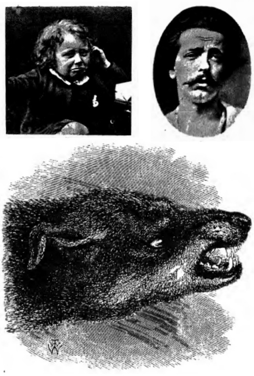
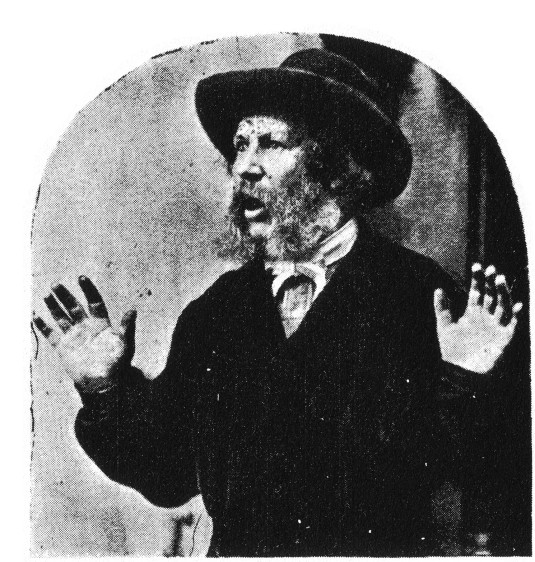
Although best known for his theory on evolution, naturalist Charles Darwin published prolifically about other topics in biology, ranging from botany, coral reefs, and even a treatise on psychology. In this 1872 text, called The Expression of Emotions in Man and Animals, Darwin suggested that similar emotional responding is found across different cultures, and to some extent, even in nonhumans. In his view, the main purpose of emotive expression is to communicate survival cues between individuals: a relaxed expression conveys safety, while a fearful expression promotes alertness, since danger may be nearby. Darwin also suggested that we gain survival information from non-human behaviors, for example, a hissing snake or a snarling lion is an immediate threat that should cause fear or other avoidance behaviors.
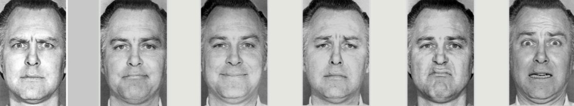
Moving forward into more modern times, the American psychologist Paul Ekman expanded on Darwin’s theory, distilling down the range of human emotions to belonging in one of seven basic categories of emotions: Anger, contempt, disgust, fear, happiness, sadness, and surprise. If the purpose of emotion was to communicate pro-survival cues, then Ekman theorized that all humans, regardless of culture, would use similar facial expressions. To test this hypothesis, Ekman visited a remote village in Papua New Guinea, where he studied a population that is isolated from any other known cultures. As predicted, these people made the same facial responses in reaction to various emotional circumstances. In 1972, Ekman published his theory of .
Part of Ekman’s research led their team to develop a series of photographs of actors portraying six major emotions. This has since been used to assess facial emotional with fascinating results: People with major depressive disorder or borderline personality disorder have a lessened ability to detect happiness in others, seeing emotional faces results in a similar emotion in the viewer, and people with dementia or Parkinson’s disease identify emotions as being less intense.
Ekman’s research led them to develop the , a system that uses facial anatomy to differentiate the features that are characteristic of different expressions. For example, a feature of a happy face is the flexing of the zygomaticus major and orbicularis oculi muscles, which produces an upward turn of the corners of the mouth and a rising of the cheeks. Other facial features, such as head movement, eye movement, and larger physical movements are also scored, and are also used to help identify emotions. The FACS can be used to formally describe why some smiles appear as genuine (a Duchenne smile, with simultaneous muscle action) while others look fake or forced (a non-Duchenne smile, characterized by the turn of the corners of the mouth without much change to the top part of the face)
15.2 Structures Involved in Emotion
Emotions are one of the most holistic functions of the brain, incorporating neural circuitry from across several different structures, ranging from the phylogenetically older to the most advanced frontal cortical areas. The structures of the Papez circuit are not a comprehensive list of brain structures involved in emotional processing but offer a good starting place for describing the anatomy of emotion.
Amygdala
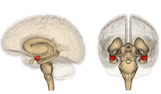
Alimbic structure called the contributes heavily to processing the valence of emotional experiences. Roughly the shape and size of an almond, the amygdala is part of the temporal lobe. Generally, the amygdala is subdivided into several nuclei, including the basolateral amygdala, central nucleus, and cortical nucleus.
As a temporal lobe structure, amygdala is strongly implicated in emotional memory formation. Emotional memories can be either positive valence (such as the happiness you may have experienced at a birthday party when younger) or negative valence (such as childhood trauma or being teased as a child).
Of the forms of declarative memory (chapter 13), autobiographical memories more often have emotional content compared to semantic memories.
Amygdala lesions have been used as a last resort treatment for patients with temporal lobe epilepsy or psychiatric conditions with pathological and dangerous aggressiveness. These psychosurgery strategies have been variably successful, but they often have high complication rates and upwards of a 4% mortality rate. These treatments are rarely used today. Deep brain stimulation may offer a less intrusive and therefore less risky therapeutic approach.
Monkeys with amygdala lesions made up the earliest descriptions of the emotional disruption condition later described as Kluver-Bucy syndrome. These monkeys did not exhibit the traditional fear response when presented with the hissing of a large snake. They also showed an absence of anger, such as when the animal is exposed to provocative novel stimuli.
A useful nonhuman model for studying emotional learning is the . In this test, a rodent is put into a cage. Occasionally, an innocuous tone and light would activate. Shortly after, an unpleasant foot-shock would be delivered to the animal. Over time, the healthy animal learns that the tone and light precede the shock to the foot, and upon future presentation of these stimuli, they will freeze. If the amygdala is lesioned, however, they freeze less, meaning they do not acquire the emotional memory associated with the stimuli. This paradigm is used as a model for post-traumatic stress disorder (PTSD), a psychiatric condition associated with the recurring recall of negative memories.
Hypothalamus
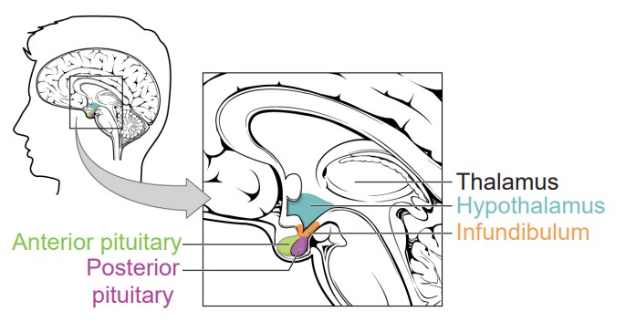
One of the major output signaling pathways of the amygdala is the , an almond-shaped nervous system structure found at the base of the brain. The hypothalamus is often seen as the neural structure that initiates endocrine responses in the rest of the body, such as hormone production. Several different behaviors, ranging from homeostasis, hunger, and circadian regulation are modulated by hypothalamic signals.
Pituitary gland
Downstream from the hypothalamus is the , a pea-sized endocrine organ that protrudes from the base of the brain. It is strongly involved in the production and release of neurohormones, signaling molecules produced by nerve cells that travel throughout the bloodstream to influence the activity of several organs throughout the entire body. The pituitary gland is subdivided into two regions with distinct anatomical and functional differences.
Posterior pituitary gland
The (also called the ) does not synthesize any neurohormones. Instead, the axonal projections of the magnocellular neurosecretory cells of the hypothalamus run through the posterior pituitary, where the hormones are secreted into a special anatomical feature called the , a series of leaky capillaries densely wrapped around the area, allowing for the hormones to easily diffuse into the bloodstream. There are predominantly two main hormones released at the posterior pituitary.
plays a significant role in the development and maintenance of , acts such as trust, compassion, and empathy, all actions that enhance interpersonal relationships. For example, increased blood levels of OT is seen in new couples compared to unattached singles, and OT release happens during orgasm, which may contribute to romantic attachment. OT signaling increases dramatically during childbirth and triggers milk letdown in lactation, which strengthens the motherchild relationship. Interestingly, while OT generally strengthens the social bonds between people, it promotes antisocial behaviors against those not perceived to be within one’s own social group.
Disorders of the OT system are believed to contribute to autism spectrum disorder and psychopathy, two complex conditions characterized partly by social impairment. Some studies have examined the therapeutic use of nasal OT for a variety of psychiatric conditions, but the studies have been unable to demonstrate strong clinical effects despite success in nonhuman animal models.
Vasopressin (AVP; or antidiuretic hormone, ADH) like OT, also contributes to social behaviors. Biochemically, AVP is very similar to OT, as they are both nine amino acid peptides differing by only two residues. AVP additionally regulates osmolarity, increasing water retention by the kidneys, which returns the body from a hypertonic state back to homeostasis. AVP also constricts blood vessels, raising blood pressure. AVP is also used therapeutically to raise blood pressure for patients in shock and to treat diabetes insipidus, a fluid dysregulation disorder (unrelated to diabetes mellitus, the much more common blood glucose disease).
Anterior pituitary
The (or the ) is capable of both synthesizing and secreting a variety of neurohormones, which contribute to several functions such as the stress response, growth, sexual development, circadian rhythms, and more. These hormones are collectively called the . The neurosecretory cells of the anterior pituitary are sensitive to signals from the hypothalamus.
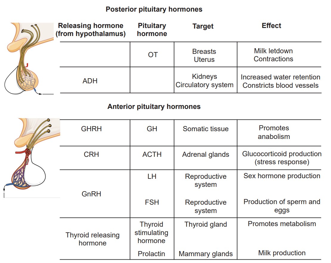
Within the anterior pituitary are cells called somatotrophs, which produce and secrete . GH is a signal that promotes , the buildup of larger molecules through biochemical reactions. Anabolic processes increase cellular repair and protein synthesis, which promote growth. Most of the time in the day, GH production is at a steady low level, but it increases at specific events, such as after meals or during slow wave sleep. The biochemical signal from the hypothalamus is .
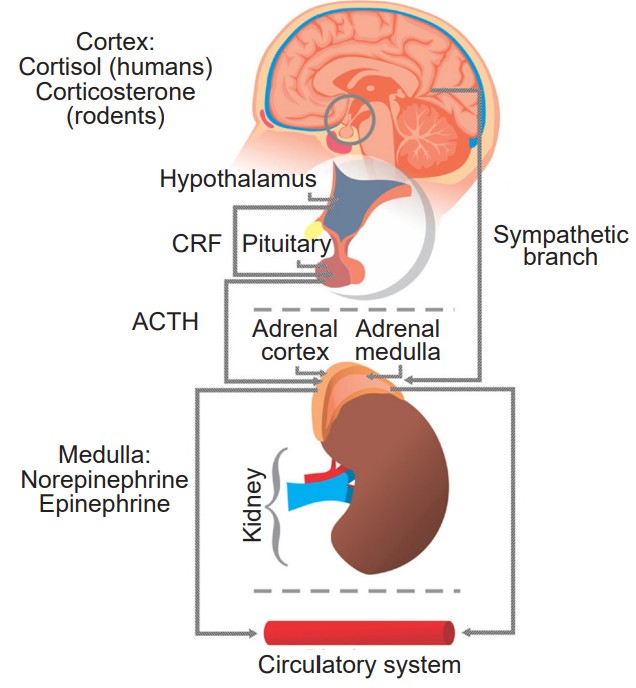
The anterior pituitary releases (or ). Once ACTH is released into the bloodstream, it travels to the , a pair of organs that sit on the anterior surface of the kidneys. Once there, ACTH triggers production of , a glucocorticoid hormone. Cortisol is best known for its initiation of the stress response, which is characterized by mild sympathetic nervous system activity. ACTH is synthesized downstream from the hypothalamic signal . The series of organs that result in the stress response is called the , or the axis for short.
The anterior pituitary also synthesizes and releases the , hormones that are important for regulation of puberty, sperm / egg production, the release of sex hormones by the testes / ovaries, and menopause. The two main gonadotropins in humans are and . These are produced in response to the hypothalamic signal . This signaling cascade is called the , or the .
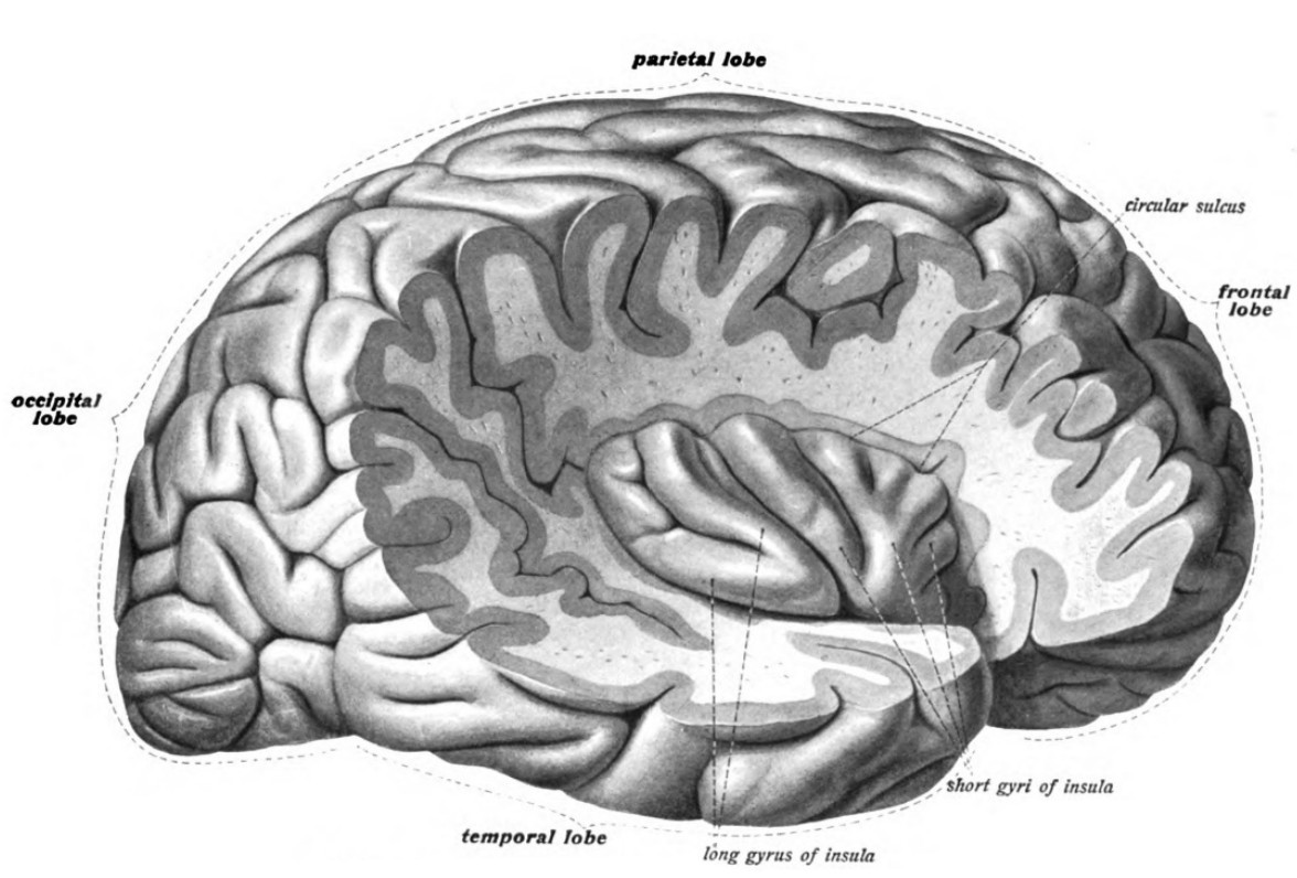
Insula
The (or ) is the lobe of the cortex buried deep within the lateral fissure. Although not visible from a side view, the insula is often considered to be the fifth lobe of the telencephalon. Early studies by Wilder Penfield, where he directly stimulated the brain during open brain surgery, led researchers to suggest that the insula contributes to , detecting the internal state of the body and conveying that information for processing.
In functional imaging studies, the insula is involved in the recall or many different emotional stimuli, especially those emotions that have a sensory component.
Notably, the insula is strongly implicated in the emotion disgust. For example, a patient is placed in an fMRI scanner while breathing through a mask, which allows the experimenters to change the smells that are perceived. Patients are then given pleasant smells (such as passion fruit, pear, or mint), a neutral smell, or unpleasant smells (like ethyl-mercaptan or isovaleric acid, which smells like skunk or body odor, respectively). There is increased activity of the anterior insula in response to the unpleasant smells but not the pleasant smells.
The insula also responds to social cues related to disgust as well. When a patient in an fMRI sees a video of a person smelling something unpleasant and reacting with a “repulsed” face (the closing of the nostrils and curling of the upper lip), their anterior insula likewise increases in activity just as if they had smelled it themselves.
In addition to sensory stimuli, feelings of social repugnance (unwarranted violence, murder) or moral disgust (incest) also increase insula activity.
Atypical insula activity is implicated in behavioral disorders. For example, insensitivity to disgust can lead to squalor-dwelling conditions (sometimes seen in excessive hoarding or late cognitive decline), which puts those people at heightened health risks due to regular exposure to unsanitary conditions. Substance use disorders, PTSD, and suicide attempts have all been associated with atypical insula activity.
15.3 Specific Emotions
Fear / Anger
Nearly everyone has experienced the prototypical fear response: Imagine reading a book when you see a spider skittering along the wall. Suddenly, you’ll feel your heart racing, your breathing increase, dryness of your mouth, and your palms sweating. You probably won’t notice the dilation of your pupils or the change in liver activity and digestion. This sympathetic nervous system activity is downstream of being presented with a fearful stimulus.
But upon closer inspection, it was no spider at all – just an errant piece of brown fuzz picked up by a draft. Within minutes, your body’s physiology returns back to normal.
This anecdote points out a few important features about the fear response. First, the onset of the fear response is quick, and so is the dissipation of the fear. Second, it is triggered by exposure to a perceived threat, regardless of whether the stimulus is a genuine threat or not (the overwhelming majority of spiders are clinically harmless to humans!). Third, the fear response is greatly modified by knowledge and experience – an entomologist would recognize that the spider is a harmless house spider and would, instead of fear, display curiosity, interest, boredom, or other emotions. On the other hand, someone who has been bitten by a dangerous spider and sent to the hospital when younger would have a much stronger physiological response.
Fear is likely the most evolutionarily ancient emotion, and is highly protective. When encountering a hungry mountain lion, faces displaying the traits of fear (enlarged eyes, flared nostrils, and a slightly open mouth accompanying a gasp) would signal to others nearby that a threat is nearby, which helps initiate heightened alertness and the appropriate fight-or-flight response.
Patient SM
is a notable case study of a person who does not experience the fear response. Born with an extremely rare genetic condition called , Patient SM progressively developed calcification in her amygdala bilaterally, causing destruction and cell death. Like other people with Urbach-Wiethe disease, she had no severe significant cognitive deficits, except for the inability to experience fear.
In one test, she was shown a variety of emotionally charged videos, then asked to rate the intensity of each clip with respect to different emotions. SM found clips from America’s Funniest Home Videos to be just as funny as the control patients, and she found the clips of disgusting toilets to be just as repulsive. But, when presented with clips depicting ghost hauntings or suspenseful serial killers on the loose, SM rated these stimuli as being non fearful.

Other studies challenged Patient SM with more concrete threats. Researchers brought SM to a pet store, where she asked to handle the snakes. She was stopped by an employee before she put her hand into a tarantula cage out of curiosity. The researchers also took her to the Waverly Hills Sanatorium haunted house, where she bravely led a group of strangers through the house populated by actors dressed as monsters and ghosts. Although she did not display any fearful behaviors, such as hesitation to walk through the darkened corridors, patient SM reported the sensation of exhilaration and enthusiasm, akin to riding a roller coaster.
She also was asked to recall some of her past real-life, fear-provoking experiences, such as when she was attacked in a domestic violence incident or was held up by a stranger at knife point in a public park. In none of these cases did she ever report feeling fear, although she was upset and angered at the situation. The destruction of her amygdala seemed to make her resilient against PTSD: the day after being threatened with a knife to her throat, she walked past the very same park bench.
Clinical connection: Plasmatoxmosis
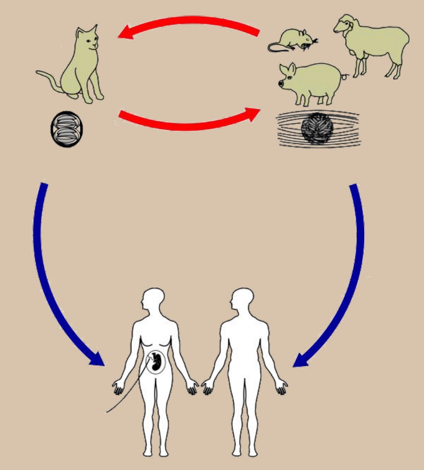
is a disease caused by infection of the parasite Toxoplasma gondii. Plasmatoxmosis may cause symptoms characteristic of a generic immune response, such as fatigue, body aches, and fever. Some people may be completely asymptomatic; however, others experience severe side effects like seizures or birth defects when an infection happens during pregnancy.
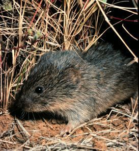
T. gondii is only capable of reproducing within cats, but the parasite has developed a very unusual evolutionary adaptation that permits propagation of the species, called the manipulation hypothesis. Cats excrete T. gondii via feces, which is eaten by rats and mice, where the parasites take up residence in their temporary host. T. gondii causes modifications in amygdala neurons, changing the rodent fear response, in turn making them attracted to cat-related odors rather than fearful of them. The fearless rodents are therefore less likely to avoid a hungry cat, increasing their risk of being eaten, which begins the cycle again.
Some research suggests that a T. gondii infection subtly affects human behavior in cat owners, hinting that T. gondii infection is associated in suicides and the onset of schizophrenia.
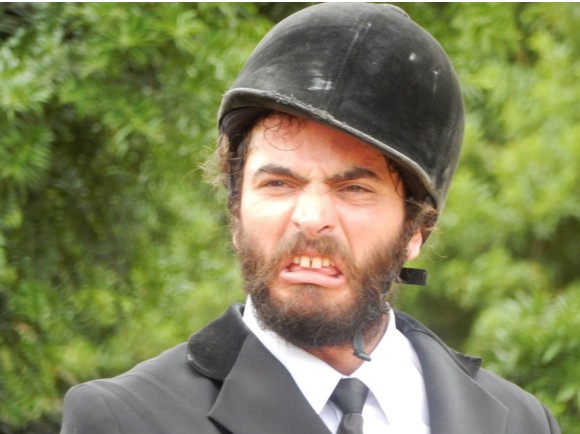
Fear and anger are two very closely related emotions. As with many other emotions, anger manifests in complex ways. The anger spectrum runs from low (irritation) to high (rage), and from quick (lashing out) to persistent (vengeful). Strong anger can provoke anti-social behavior such as violence.
Since anger may precede violence, angry faces function as a warning during evolutionary survival situations or during interpersonal conflict. Seeing angry faces may prompt awareness, initiating the sympathetic nervous system response in case “fight or flight” becomes necessary. As with fear, the emotional response to angry faces or body postures is partially guided by the amygdala, resulting in communication with the rest of the body through cortisol release downstream of the HPA axis.
Environmental factors such as childhood maltreatment or expectations are partially responsible for how a person responds to a particular anger-provoking stimulus. Internal homeostatic conditions also influence the anger response, just as how low blood sugar increases aggression and drives negative or hateful feelings in the face of a challenge (the portmanteau “hangry” was added to the Oxford Dictionary in 2018).
Neurobiological factors also contribute to the anger response. In addition to amygdala circuits, regions in the frontal cortex decrease activity during acts of aggression, suggesting that frontal circuits actively inhibit the limbic system, which drive our more “primitive” responses. Altered frontal cortical action may therefore account for one reason why two different people would react to the same anger-provoking stimulus in different ways.
Clinical connection: Borderline Personality Disorder (BPD)
is an emotional dysregulation disorder characterized by disproportionately intense emotional reactivity to environmental triggers. For instance, they may experience anger or hostility in response to minor inconveniences. These emotional changes may persist for unusually long durations, up to hours or days, which often puts strain on interpersonal relationships. They also experience a fear of abandonment, an unstable sense of self, suicidal ideation or attempts, and dissociation (an emotional detachment from events).
It is estimated that 1.4% of people have BPD. It is often comorbid with other psychiatric disorders, such as anxiety, depression, or substance use disorders. The main treatment approach is psychotherapy; medications are not always effective at reversing the symptoms.
Celebrities such as Saturday Night Live actor Pete Davidson, musician Amy Winehouse, and Princess Diana likely had BPD. Arguably, the fictional character Anakin Skywalker from the Star Wars prequel films also displays many diagnostic traits of BPD.
Stress
Stress is a natural and healthy bodily response that is similar to the prototypical “fightor-flight” response: elevated heart rate and blood pressure, increased respiration rate (shortness of breath), energy usage, and perspiration, among others. Physiologically, stress also causes a weakening of the immune response, a slowing of growth, and decreases in kidney filtration.
Stress can either result from purely physical stimuli (exposure to constant cold; chronic dehydration), a purely psychological threat (resentment; religious guilt), or a combination of both (living paycheck to paycheck).
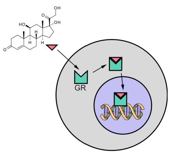
The human stress hormone cortisol triggers the physiological response to stress. is a signaling molecule synthesized from cholesterol which readily passes through cell membranes where it can activate intracellular . Once the ligand-receptor complex is formed, it translocates into the nucleus, where it functions as a transcriptional regulator. Nearly every cell contains GRs, meaning that an elevation of systemic cortisol can influence several organs to change their activity.
A small amount of stress, sometimes called , is beneficial and generally performance-enhancing, such as in the moments leading up to the beginning of an athletic competition. The opposite, , is characterized by the inability to cope with rising demands, leading to increased anxiety and other maladaptive responses. Distress is the kind of stress that people experience when they are facing multiple rapidly-approaching deadlines, difficulties in paying rent, or relationship issues.
Biochemically and physiologically, eustress is the same as distress. The major difference is the psychological interpretation or perception of the stressor. Eustress is seen as a challenge which someone can work to overcome and is often short lived or temporary. Chronic long-term distress causes all variety of negative health outcomes, such as cardiovascular disease, diabetes, and obesity, all risk factors for premature death.
The early medical descriptions of stress were put forth by Hans Selye in the 1930’s. As a medical student, he observed that chronically ill humans, regardless of their specific condition, often experienced a similar set of late-stage negative health outcomes, such as gastric ulcers, high blood pressure, and heart attacks. Collectively, he called this response the .
Selye observed that extracts from different internal organs led to these health changes in rats, eventually leading to premature death. As a good scientist, Selye also injected a second group of rats with a harmless saline solution as a control. Surprisingly, Selye observed that these control animals also exhibited the general adaptation syndrome.
Clinical connection: Ulcers
are injuries to the mucous membranes of the stomach or intestines resulting in abdominal pain, nausea, and changes in appetite (although some patients report no symptoms.) In severe cases, ulcers can cause bleeding or perforation of the stomach, which could be fatal. An estimated 4% of people have ulcers, although this is likely an underestimation.
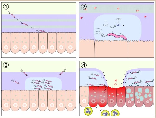
For several years, ulcers were believed to be caused solely by stress. However, later research suggested that colonies of the gut bacterium Helicobacter pylori was found to contribute to the formation of 80% of ulcers, a discovery that earned Drs. Barry Marshall and Robin Warren a Nobel Prize in 2005. Today, the evidence suggests that neither factor exclusively leads to ulcer formation: Nearly half of all people have the H. pylori colonies in their gut, but not all these people develop ulcers. Also, about 30% of people with ulcers do not harbor H. pylori, and people taking oral antibiotics that have eliminated their H. pylori colonies can still develop ulcers.
It turns out that even minor stressors, such as the movement of the rat cage, grabbing of the animal, and administering an injection led to an eightfold increase in plasma levels of epinephrine, a hormone upstream of the stress response. Selye concluded that the general adaptation syndrome was not a result of the injections of organ extract, but rather because he induced a tremendous stressor to all the rats.
Like Selye, we use nonhuman models to study how the nervous system changes in response to stress. Rodents can be exposed to , where they are put into a narrow cylinder for part of their day. is a rodent model of starvation or dietary malnutrition. The is best known as an assessment of depressive behavior, but it can also be used to induce stress. Food restriction and the forced swim test increases plasma levels of the neurohormone (the rodent analog of cortisol).
Psychological stressors also induce the stress response. Common stress paradigms include exposure to socially-dominant bully rats or predator-associated cues like fox urine, which contains a group of chemicals called kairomones. cause behavioral changes in other species, whereas pheromones refer to interspecies chemical communication. Social isolation is also a stressor for rodents, and for this reason, rats and mice are normally group housed.
Love
Love can be thought of as an intensely strong attachment towards a person (romantic love, lust), a thing (passion project), or concept (patriotism as a love for country, or altruism as a love for fellow humans). However, it is very difficult to put a strict biological definition on these varied concepts of love.
This section focuses on interpersonal love, of which there are several unique forms, each resulting in different behavioral outcomes. For example, romantic love drives physical attraction, lust, and sexual activity. Parental love, on the other hand, encourages self-sacrifice and hyper-attentiveness towards a newborn. Some behaviors are shared between the two forms of love, such as respect.
Romantic love
Romantic love drives much of human behavior and has been documented thoroughly in the arts all the way from Homer’s Iliad through Shakespeare’s Romeo and Juliet up to modern Taylor Swift.

The majority of human societies have embraced , the romantic relationship characterized by a pair of people who share resources, parenting duties, and exhibit preferential mating. Outside of humans however, only 9% of mammalian species form socially monogamous pairs, while at least 75% of bird species maintain socially monogamous relationships (which may explain the origin of the phrase “love birds”).
Dr. Helen Fisher, an anthropologist and a leader in the field of love research, suggests that love can be divided into three closely interconnected components. These three are guided in part by different signaling pathways, and lead to somewhat different behavioral outcomes.
1. Lust
(or ) refers to a very strong desire for sexual gratification. These behaviors are largely driven by the actions of the sex hormones , , and , released downstream of activation of the HPG axis. As hormones, they are synthesized from cholesterol and circulate through the bloodstream to influence the body in many ways.
Both testosterone and estradiol contribute to sex-seeking behaviors in men and women, where increasing testosterone levels drive up sexual desire.
In the brain, the sex hormones strongly influence the of the anterior hypothalamus. The mPOA contains a , the part of the brain that exhibits the biggest morphological difference between males and females: in humans, it is about twice as large in males with double the number of neurons throughout childhood and early adulthood. In the rat, it is up to 8 times larger in males than females, and if this area is lesioned, rats exhibit decreased motivation to engage in sex.
The amygdala also plays a significant role in mediating lust, and lesions may either result in hypersexuality (Kluver-Bucy syndrome) or a decrease in responding to socially-derived sex cues.
In addition to changing libido, the sex hormones contribute to sexual differentiation and maturation (mascularization with testosterone, such as muscle mass increases and hair growth), differences in immune system activity (decreases with higher testosterone), and pain tolerance (higher tolerance with higher testosterone) for example. Progesterone is less of a behavioral driver and functions more as a signal that prepares female bodies for changes in preparation for childbirth, such as in the uterus and breasts.
2. Attraction
is characterized by high energy investment and preoccupation towards a small number of people. From an evolutionary perspective, attraction may have developed to discriminate between multiple reproductive partners, allowing the focusing of limited resources towards fewer partners.
In Fisher’s theory, attraction is strongly related to the action of dopamine and norepinephrine. In humans, the reward circuitry (Section 11.2) is involved in feelings of love. Using fMRI, Fisher presented pictures of a patient’s romantic partner to them and identified increases in the blood flow to dopaminergic midbrain areas such as the ventral tegmental area and the striatum. This finding compares with imaging studies that observed increases in blood flow to insula, premotor, and hypothalamus as well as striatum in response to highly erotic, sexual imagery. These studies suggest that romantic love and lust have different driving neural structures underlying these behaviors.
Norepinephrine functions to increase attention, alertness, and energy, which accounts for the exhilarated feeling you may feel when spending time with a potential partner.
3. Attachment
is the long-term accompanied by feelings of comfort and emotional stability. Attachment also contributes to behaviors that maximize offspring survivability, such as sharing parenthood responsibilities and protectiveness towards offspring. The major neurochemical drivers of this form of love are oxytocin and vasopressin.
We have learned about mammalian pair bonding through experiments with (Microtus ochrogaster), rodents that are indigenous to central North America. Atypical of mammals, prairie voles exhibit social monogamy after mating, where the two voles will cohabitate and share parental duties, such as mutual resource collection, nest building, and caring for their young.
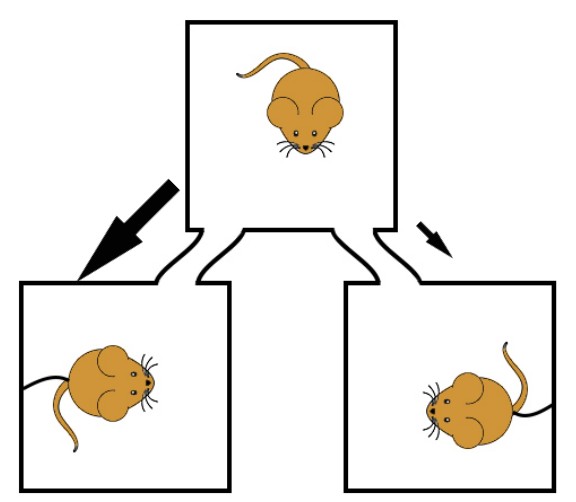
An experimental test called the can be used to assess vole monogamy. Here, a three-chamber testing apparatus is used where a test vole is placed in a central chamber. In the other two chambers are voles who have been harnessed into their rooms, unable to leave. The test vole is then free to move between any of the three chambers. When the test vole has mated with one of the harnessed voles, they will choose to spend more time with their “partner” vole compared to the novel “stranger” vole.
Not only do mated prairie voles prefer the company of their partner, they also demonstrate behaviors similar to some human romantic relationships. For example, mated prairie voles living together display selective aggression against a stranger, “intruder” voles, a behavior called “mate-guarding”. They also spend significant time mutually caring for their young, their pair bonding can be modified by psychoactive substance use, and they show increased anxiety when they are separated from their partner vole.
Other nonhuman studies have compared the prairie voles with physiologically comparable animal models, such as the montane vole or meadow voles. While these other rodents are similar in size, they have slightly different geographic distributions. Notably, they have radically different mating habits, as they exhibit social promiscuity rather than monogamy.
Comparing between monogamous and promiscuous species has allowed researchers to identify several neurotransmitter signals that are implicated in pair bonding. Oxytocin and vasopressin seem to play important roles in vole pair bonding, as inhibition of either of these signals decrease partner preference. Additionally, differences in dopamine receptor levels and corticotropin releasing factors are observed between vole species.
Parental love
Parental love refers to instinctive affection towards one’s offspring. Parental love behaviors include nurturing (collecting and sharing resources), protecting (promoting aggression against “intruders”), and preparing one’s young for their adult life (risk assessment training).
In evolutionary theory, parental love serves to improve the odds of passing of one’s genes through the following generation. Most large mammals (humans included) are , which benefit most from a small number of high-quality offspring that require substantial parental investment, as opposed to large numbers of offspring with little investment.
An extension of parental love is or , the protection and preferential support of one’s extended genetic relations. Because blood relatives share common genes, increasing the survivability of these family members has benefits for passing genes to the next generation. In the words of the geneticist Jack Haldane, “I’d gladly give my life for two brothers or eight cousins.”
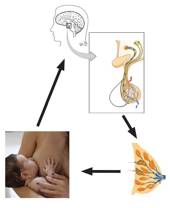
Many behaviors related to mammalian motherhood are accompanied by changes in neural activity. Nursing, for instance, is feeding behavior that is regulated through a positive feedback cycle. Offspring suckling activates the mother’s somatosensory afferents. Through a series of oxytocin-dependent circuits across the hypothalamus, suckling ultimately increases lactation through the . Auditory sensory inputs such as the sounds of a crying baby can also trigger this reflex. Sometimes, just thinking about the baby can induce letdown.
Some changes are dependent on neural plasticity. For example, after childbirth, the auditory areas of rodents rewire to become more sensitive to high frequency sounds. This adaptation allows the mothers to better detect the that are emitted by offspring when they are distressed or hungry. Olfactory areas also change in order to become more sensitive to the particular odorants given off by their young, allowing them to better identify their offspring. In humans, these olfactory changes result in decreased aversion towards traditionally aversive stimuli (urine or fecal matter) when they originate from their children.
Disgust
Disgust is an evolutionarily protective emotion that helps protect an organism from being exposed to toxic or dangerous substances. It also leads to disease avoidance, which is why we dislike the smell of rotten foods or fecal matter, and why we may be repulsed by blood. As in the previous examples, disgust often happens in response to specific sensory stimuli (chapter 9).
The most common physiological response to disgusting stimuli is nausea, which can promote vomiting. Additionally, nausea decreases appetite, which prevents further intake of potentially toxic compounds.
Disgust is not inherently ingrained and has a strong learned cultural component that can be modified by experience. Americans may be turned off by potent moldy cheeses, which are delicacies in many European countries, or fermented fish products, commonly used as a flavor enhancer in South East Asian cuisine.
Disgust extends beyond physical or chemical stimuli, and also manifests itself in thought. For instance, disgust is associated with sexual contact leading to low reproductive success, such as with animals or incestuous relationships. Antisocial behaviors can also provoke the disgust behavior, such as feeling physically nauseated when reading about serial killers.
The canonical “disgust face” is observed across several cultures. The curling of the upper lip and wrinkling of the nose decrease air flow through the nasal cavity, and squinting minimizes eye surface exposure. An opening of the mouth is a precursor for spitting out the disgusting flavors.
The insula is involved in mediating perception of disgust and displays changes in activity in fMRI activity in response to disgust-related cues.
Clinical connection: Obsessive-compulsive disorder (OCD)
OCD is a common psychiatric condition affecting an estimated 3% of the population, characterized by persistent intrusive thoughts or the need to perform some ritualistic series of actions or motions. OCD exists on a spectrum, and in severe cases, can severely impair the quality of a patient’s life. OCD is often treated with psychiatric intervention or SSRIs like fluoxetine (more information in chapter 16)
Some studies implicate abnormal disgust sensitivity as contributing to OCD. Patients were shown a variety of disgust-provoking stimuli, such as unsanitary toilets or parasitic infections. If the threshold of disgust sensation is particularly low a patient may engage in their perseverative behavior more frequently
Image credits
15.1 https://commons.wikimedia.org/wiki/File:The_Feeling_Wheel.png
15.3 https://pixabay.com/photos/bicycle-bike-biking-sport-cycle-384566/
15.4 https://pixabay.com/photos/cat-cat-hissing-angry-pet-5534007/
15.5 https://commons.wikimedia.org/wiki/File:Papez.jpg modified by Austin Lim
15.6 https://pixabay.com/photos/lion-tailed-macaque-monkey-rare-216227/
15.8 https://upload.wikimedia.org/wikipedia/commons/5/5a/Expression_Of_The_Emotions_In_Man_And_Animals_%28IA_
TheExpressionOfTheEmotionsInManAndAnimals%29.pdf
15.9 https://commons.wikimedia.org/wiki/File:Facial_Action_Coding_System_-_Paul_Ekman_%26_Wallace_Friesen.png
15.10 https://commons.wikimedia.org/wiki/File:Amygdala.png
15.11 https://commons.wikimedia.org/wiki/File:1806_The_Hypothalamus-Pituitary_Complex.jpg
15.12 https://upload.wikimedia.org/wikipedia/commons/4/4a/1810_Major_Pituitary_Hormones.jpg modified by Austin Lim
15.13 https://upload.wikimedia.org/wikipedia/commons/b/b4/Sobo_1909_633.png
15.14 https://commons.wikimedia.org/wiki/File:Expression_of_the_Emotions_Plate_VII_(fear1).jpg 15.15 https://commons.
wikimedia.org/wiki/File:Gigi_a_motley_yellow_corn_snake.jpg https://upload.wikimedia.org/wikipedia/commons/e/eb/
Waverlyhillssanatorium.jpg
15.16 https://phil.cdc.gov/Details.aspx?pid=3421 modified by Austin Lim
15.17 https://commons.wikimedia.org/wiki/File:Response_to_stress.jpg modified by Austin Lim
15.18 https://commons.wikimedia.org/wiki/File:DNA_simple2-bn.svg modified by Austin Lim
https://commons.wikimedia.org/wiki/File:Cortisol.png
15.19 https://commons.wikimedia.org/wiki/File:H_pylori_ulcer_diagram.png
15.20 https://commons.wikimedia.org/wiki/File:Two_swans_in_a_lake_(23746714432).jpg
15.21 https://commons.wikimedia.org/wiki/File:Prairie_vole.gif
15.22 https://pixabay.com/vectors/mouse-rodent-gnawer-white-animal-146416/ modified by Austin Lim
15.23 https://commons.wikimedia.org/wiki/File:2922_Let_Down_Reflex-new.jpg modified by Austin Lim https://pixabay.
com/photos/breastfeeding-mother-mother-s-love-1570695/ modified by Austin Lim
15.24 https://commons.wikimedia.org/wiki/File:How_to_express_disgust_(33491312650).jpg
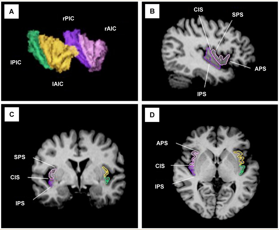
15.25 Disproportionate Alterations in the Anterior and Posterior Insular Cortices in Obsessive–Compulsive Disorder. Song
A, Jung WH, Jang JH, Kim E, Shim G, et al. (2011) Disproportionate Alterations in the Anterior and Posterior Insular Cortices
in Obsessive–Compulsive Disorder. PLOS ONE 6(7): e22361. https://doi.org/10.1371/journal.pone.0022361
field studying neural mechanisms that underlie emotion
says the body’s physiological changes precede the onset of an emotional response
serves as a communication route to the body’s endocrine system
handles sensory information
procedure where the cortex is surgically separated from the rest of the nervous system
hyper-aggressive responses to stimuli
certain structures in the brain that have a role in emotional behavior
the inability to recognize faces or objects visually
an inappropriate fixation with using the mouth to interact with surroundings, such as licking or eating nonfoods
a unique set of emotional deficits causing a failure to display fear or anger, visual agnosia, psychic blindness, hypersexuality, and hyperorality
says people use a combination of the physiological response and a cognitive label to determine the emotion that is most appropriate for a given circumstance
Paul Ekman's theory that all humans, regardless of culture, would use similar facial expressions to communicate the basic categories of emotions
neuropsychological tool assessing emotion recognition from facial expressions
a system that uses facial anatomy to differentiate the features that are characteristic of different expressions
brain structure contributing heavily to processing valence of emotional experiences
nonhuman model for studying emotional learning
strongly involved in the production and release of neurohormones
one of two lobes in the pituitary gland; produces oxytocin and vasopressin
another name for Posterior pituitary gland
a series of leaky capillaries densely wrapped around the posterior pituitary
plays a significant role in the development and maintenance of prosocial behaviors
acts such as trust, compassion, and empathy, all actions that enhance interpersonal relationships
one of two halves of the Pituitary gland capable of both synthesizing and secreting a variety of neurohormones
another term for anterior pituitary
collection of hormones responsible for several functions such as the stress response, growth, sexual development, circadian rhythms, and more
enhances cellular repair, muscle and bone growth, and protein synthesis
the buildup of larger molecules through biochemical reactions
biochemical signal from the hypothalamus
released by the anterior pituitary and triggers production of cortisol
short for adrenocorticotropic hormone
a pair of organs that sit on the anterior surface of the kidneys
a glucocorticoid hormone best known for its initiation of the stress response
drives the bodies response to stress
series of organs that result in the stress response
short for hypothalamic-pituitary-adrenal axis
hormones that are important for regulation of puberty, sperm or egg production, the release of sex hormones by the testes or ovaries, and menopause
one of two main gonadotropins in humans
one of two main gonadotropins in humans
signals production of the two main gonadotropins in humans: the luteinizing hormone (LH) and the follicle-stimulating hormone (FSH)
signaling cascade involving the production of gonadotropins in the Pituitary gland
short for hypothalamic-pituitary-gonadal axis
the lobe of the cortex buried deep within the lateral fissure
another term for the insula
detecting the internal state of the body and conveying that information for processing
a notable case study of a person who does not experience the fear response
condition causing a calcification in the amygdala bilaterally leading to the inability to experience fear
disease caused by infection of the parasite Toxoplasma gondii; may cause symptoms characteristic of a generic immune response, such as fatigue, body aches, and fever
an emotional dysregulation disorder characterized by disproportionately intense emotional reactivity to environmental triggers
the receptor in which cortisol binds to; contained in nearly every cell
a small amount of stress that is generally beneficial and performance enhancing
the inability to cope with rising demands, leading to increased anxiety and other maladaptive responses
a collection of responses observed by Hand Selye in chronically ill humans, including gastric ulcers, high blood pressure, and heart attacks
injuries to the mucous membranes of the stomach or intestines resulting in abdominal pain, nausea, and changes in appetite
method used to induce physiological responses in an animal by restricting its free movement
a rodent model of starvation or dietary malnutrition
an assessment of depressive behavior, and can also be used to induce stress
the rodent analog of cortisol
q group of chemicals that cause behavioral changes in species
the romantic relationship characterized by a pair of people who share resources, parenting duties, and exhibit preferential mating
a very strong desire for sexual gratification
another term for lust
sex hormone in men
sex hormone in women
known as the pregnancy hormone in women
section of the anterior hypothalamus in the brain strongly influenced by sex hormones
the part of the brain that exhibits the biggest morphological difference between males and females (twice as large in men)
characterized by high energy investment and preoccupation towards a small number of people
the long-term accompanied by feelings of comfort and emotional stability
rodents that are indigenous to central North America
an experimental test used to assess vole monogamy
species that benefit from a few high-quality offspring rather than a large number of offspring with little investment
the protection and preferential support of one’s extended genetic relations
another term for familial love
a natural process in mammalian motherhood where offspring suckling activates a lactation cycle through oxytocin-dependent circuits
sounds emitted by rodent offspring when they are distressed or hungry
