
4.3 Transport Mechanisms Used for Uptake and Absorption
Transport Mechanisms Used for Uptake and Absorption
There are a number of different transport mechanisms utilized by your body for the uptake of nutrients into cells, and absorption into the bloodstream. These mechanisms can be classified as being either passive transport or active transport. The difference between the two types of transport is whether energy is required, and whether they move with or against a concentration gradient. Passive transport does not require energy and moves with a concentration gradient (high to low concentration). Active transport requires energy to move against the concentration gradient (low to high concentration).
A concentration gradient is a result of an unequal distribution of solutes within a solution. A solute is what is dissolved in a solvent in a solution. The more solute a region has, the higher the its concentration, while the less solute a region has, the lower the its concentration. Moving with the gradient is moving from a region of higher concentration to an area of lower concentration. Moving against the gradient is moving from an area of lower concentration to an area of higher concentration.

Figure 4.31 Movement with and against a concentration gradient.
Because our cells are surrounded by fluids containing varying amounts of solute, our body cells can experience concentration gradients across the plasma membrane. Hypertonic refers to a situation when the cell is surrounded by a solution that contains more solute than inside the cell. Hypotonic refers to a situation when the cell is surrounded by a solution containing less solutes than inside the cell. Isotonic refers to a situation when the cell is surrounded by a solution containing the same number of solutes that inside the cell. Figure 4.32 demonstrates these well.


Figure 4.32 Tonicity across the plasma membrane of cells.
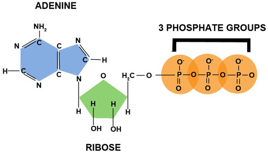
The energy for active transport is provided by adenosine triphosphate (ATP), which is the energy currency in the body. Tri- means three, thus ATP is adenosine (composed of an adenine and a ribose) with three phosphate groups bonded to it, as shown below.

Figure 4.33 Structure of adenosine triphosphate (ATP)1
Phosphorylation is the formation of a phosphate bond. Dephosphorylation is the removal of a phosphate bond. Phosphorylation is an anabolic process that requires energy.
Dephosphorylation is a catabolic process that releases energy. Thus, energy is required to add phosphates to ATP, while energy is released through removing phosphates from ATP. Figure
- depicts this process.

Figure 4.34 The ATP Cycle demonstrating the processes of phosphorylation and dephosphorylation.
Subsections:
- 4.31 Passive Transport Mechanisms
- 4.32 Active Transport Mechanisms
References & Links
1. https://userscontent2.emaze.com/images/92312f55-f2da-4d80-a1c3- 657851b8e450/bf6e4c6e-7a39-406f-aac7-b422321028a5.jpg
Passive Transport Mechanisms
There are three forms of passive transport involved in uptake and absorption of nutrients in the body:
- Simple Diffusion
- Osmosis
- Facilitated Diffusion
Simple Diffusion
Simple diffusion is the movement of solutes from an area of higher concentration to an area of lower concentration (with the concentration gradient) without the help of a protein, as shown
in Figure 4.311.

Figure 4.311 Simple diffusion
Osmosis
Osmosis is similar to simple diffusion, but water moves instead of solutes. In osmosis water molecules move from an area of lower solute concentration to an area of higher solute concentration as shown below. The effect of this movement is to dilute the area of higher concentration.

Figure 4.312 Osmosis
The following videos do a nice job of illustrating osmosis.
Required Web LinksVideo: Osmosis (0:47)Video: Osmosis in the Kitchen (0:58)
Another example illustrating osmosis is the red blood cells in different solutions shown below.

Figure 4.313 Effect of salt solution concentration on red blood cells1
We will consider the simple example of salt as the solute. If the solution is hypertonic, that means that there is a greater concentration of salt outside (extracellular) the red blood cells than within them (intracellular). Water will then move out of the red blood cells to the area of higher salt concentration, resulting in the shriveled red blood cells depicted. Isotonic means that there is no difference between concentrations. There is an equal exchange of water between intracellular and extracellular fluids. Thus, the cells are normal, functioning red blood cells. A hypotonic solution contains a lower extracellular concentration of salt than the red blood cell intracellular fluid. As a result, water enters the red blood cells, possibly causing them to burst.
Facilitated Diffusion
The last form of passive transport is similar to diffusion in that it also moves with the concentration gradient (higher concentration to lower concentration). While it requires no energy, it does require a carrier protein to transport the solute across the membrane. Figure
4.314 and Required Video Link do a nice job of illustrating facilitated diffusion.

Figure 4.314 Facilitated diffusion examples2
Required Web LinkVideo: Facilitated Diffusion (0:27)
References & Links
1. http://en.wikipedia.org/wiki/File:Osmotic_pressure_on_blood_cells_diagram.svg 2.https://en.wikipedia.org/wiki/Facilitated_diffusion#/media/File:Scheme_facilitated_diffusion
_in_cell_membrane-en.svg
Videos
Osmosis – http://www.youtube.com/watch?v=sdiJtDRJQEc
Osmosis in the Kitchen – http://www.youtube.com/watch?v=H6N1IiJTmnc&NR=1&feature=fvwp
Facilitated Diffusion – http://www.youtube.com/watch?v=s0p1ztrbXPY
Active Transport Mechanisms
There are two forms of active transport:
- Active Carrier Transport
- Endocytosis
Active Carrier Transport
Active carrier transport (sometimes referred to as secondary active transport) is similar to facilitated diffusion in that it utilizes a protein carrier. However, energy is also required to move compounds against their concentration gradient (lower to higher concentration). Figure 4.321 and video do a nice job of illustrating active carrier transport.

Figure 4.321 Sodium-potassium ATPase (aka sodium-potassium pump) an example of active carrier transport1
Required Web LinkVideo: Active Transport (0:21)
Endocytosis
Endocytosis is the engulfing of particles, or fluids, to be taken up into the cell. If a particle is endocytosed, this process is referred to as phagocytosis. If a fluid is endocytosed, this process is referred to as pinocytosis. Whenever a receptor located on the membrane is used to assist in engulfing an extracellular component, it is known as receptor mediated endocytosis. These processes are shown in Figure 4.322.

Figure 4.322 Different types of endocytosis2
The following video does a really nice job of showing how endocytosis occurs.
Required Web LinkVideo: Endocytosis (0:35)
References & Links
- https://en.wikipedia.org/wiki/File:Scheme_sodium-potassium_pump-en.svg
- http://commons.wikimedia.org/wiki/File:Endocytosis_types.svg
Videos
Active Transport – http://www.youtube.com/watch?v=STzOiRqzzL4 Endocytosis – http://www.youtube.com/watch?v=4gLtk8Yc1Zc
